

Epithelial-mesenchymal transition in cancer metastasis through the lymphatic system
- PMID: 28590032
- PMCID: PMC5496496
- DOI: 10.1002/1878-0261.12092
Epithelial-mesenchymal transition in cancer metastasis through the lymphatic system
Abstract
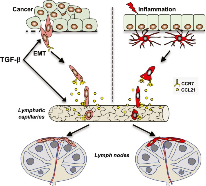
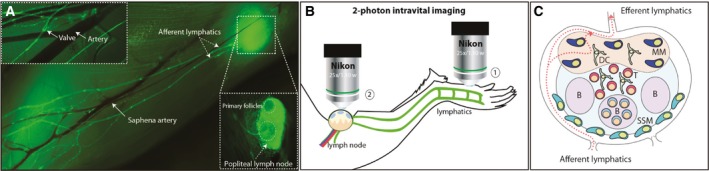
It was already in the 18th century when the French surgeon LeDran first noted that breast cancer patients with spread of tumor cells to their axillary lymph nodes had a drastically worse prognosis than patients without spread (LeDran et al., ). Since then, metastatic spread of cancer cells to regional lymph nodes has been established as the most important prognostic factor in many types of cancer (Carter et al., ; Elston and Ellis, ). However, despite its clinical importance, lymph metastasis remains an underexplored area of tumor biology. Fundamental questions, such as when, how, and perhaps most importantly, why tumor cells disseminate through the lymphatic system, remain largely unanswered. Accordingly, no treatment strategies exist that specifically target lymph metastasis. The identification of epithelial-mesenchymal transition (EMT) as a mechanism, which allows cancer cells to dedifferentiate and acquire enhanced migratory and invasive properties, has been a game changer in cancer research. Conceptually, EMT provides an explanation for why epithelial cancers with poor differentiation status are generally more aggressive and prone to metastasize than more differentiated cancers. Inflammatory cytokines, such as TGF-β, which are produced and secreted by tumor-infiltrating immune cells, are potent inducers of EMT. Thus, reactivation of EMT also links cancer-related inflammation to invasive and metastatic disease. Recently, we found that breast cancer cells undergoing TGF-β-induced EMT acquire properties of immune cells allowing them to disseminate in a targeted fashion through the lymphatic system similar to activated dendritic cells during inflammation. Here, we review our current understanding of the mechanisms by which cancer cells spread through the lymphatic system and the links to inflammation and the immune system. We also emphasize how imaging techniques have the potential to further expand our knowledge of the mechanisms of lymph metastasis, and how lymph nodes serve as an interface between cancer and the immune system.
Keywords: EMT; TGF-β; chemokines; immune cell properties; lymph metastasis; targeted migration.
© 2017 The Authors. Published by FEBS Press and John Wiley & Sons Ltd.
Figures
References
-
- Allard WJ, Matera J, Miller MC, Repollet M, Connelly MC, Rao C, Tibbe AGJ, Uhr JW and Terstappen L (2004) Tumor cells circulate in the peripheral blood of all major carcinomas but not in healthy subjects or patients with nonmalignant diseases. Clin Cancer Res 10, 6897–6904. - PubMed
-
- Ansieau S, Bastid J, Doreau A, Morel AP, Bouchet BP, Thomas C, Fauvet F, Puisieux I, Doglioni C, Piccinin S et al (2008) Induction of emt by twist proteins as a collateral effect of tumor‐promoting inactivation of premature senescence. Cancer Cell 14, 79–89. - PubMed
-
- Baluk P, Hashizume H and McDonald DM (2005) Cellular abnormalities of blood vessels as targets in cancer. Curr Opin Genet Dev 15, 102–111. - PubMed
-
- Bertran E, Caja L, Navarro E, Sancho P, Mainez J, Murillo MM, Vinyals A, Fabra A and Fabregat I (2009) Role of cxcr4/sdf‐1 alpha in the migratory phenotype of hepatoma cells that have undergone epithelial‐mesenchymal transition in response to the transforming growth factor‐beta. Cell Signal 21, 1595–1606. - PubMed
Publication types
MeSH terms
Substances
LinkOut - more resources
Full Text Sources
Other Literature Sources
