

Monitoring the Retention of Human Proliferating Cell Nuclear Antigen at Primer/Template Junctions by Proteins That Bind Single-Stranded DNA
- PMID: 28590137
- PMCID: PMC5662943
- DOI: 10.1021/acs.biochem.7b00386
Monitoring the Retention of Human Proliferating Cell Nuclear Antigen at Primer/Template Junctions by Proteins That Bind Single-Stranded DNA
Abstract
In humans, proliferating cell nuclear antigen (PCNA) sliding clamps encircling DNA coordinate various aspects of DNA metabolism throughout the cell cycle. A critical aspect of this is restricting PCNA to the vicinity of its DNA target site. For example, PCNA must be maintained at or near primer/template (P/T) junctions during DNA synthesis. With a diverse array of cellular factors implicated, many of which interact with PCNA, DNA, or both, it is unknown how this critical feat is achieved. Furthermore, current biochemical assays that examine the retention of PCNA near P/T junctions are inefficient, discontinuous, and qualitative and significantly deviate from physiologically relevant conditions. To overcome these challenges and limitations, we recently developed a novel and convenient Förster resonance energy transfer (FRET) assay that directly and continuously monitors the retention of human PCNA at a P/T junction. Here we describe in detail the design, methodology, interpretation, and limitations of this quantitative FRET assay using the single-stranded DNA-binding protein, SSB, from Escherichia coli as an example. This powerful tool is broadly applicable to any single-stranded DNA-binding protein and may be utilized and/or expanded upon to dissect DNA metabolic pathways that are dependent upon PCNA.
Figures
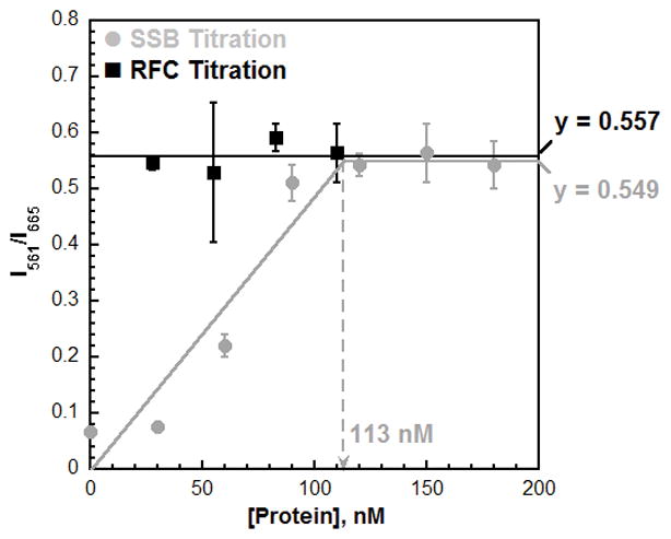
 ) or saturated with SSB (180 nM) and titrated with RFC (30 – 110 nM) (·). Results are plotted versus the concentration of the respective titrant. When SSB was titrated, the FRET signal increased linearly with SSB concentration up to 90 nM. Data points within this concentration range (0 – 90 nM SSB) were fit to a linear equation. After 90 nM, the FRET plateaus and remains constant with SSB concentration. Data points within this concentration range (120 – 180 nM) were fit to a flat line. The two lines intersect at the “breakpoint” where the Cy3P/BioT70 DNA substrate is saturated with Cy5-PCNA. When RFC was titrated, the FRET signal remained constant at a level (0.557) equivalent to that observed at saturating concentrations of RFC and SSB (0.549). These FRET values are identical to those observed for RPA (Table S1). This suggests that Cy5-PCNA encircling a P/T junction is in the same FRET state when the adjacent ssDNA is bound by either RPA or SSB.
References
-
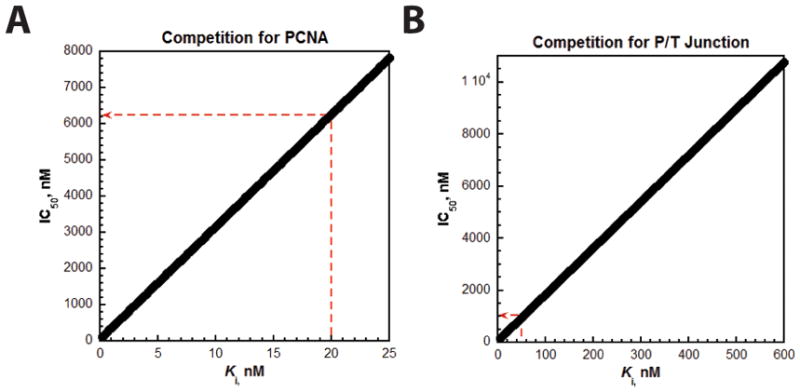
- Munson PJ, Rodbard D. J Recept Res. 1988;8:533–546. - PubMed
Publication types
MeSH terms
Substances
Grants and funding
LinkOut - more resources
Full Text Sources
Other Literature Sources
Miscellaneous
