. 2017 Aug 3;16(15):1404-1413.
doi: 10.1080/15384101.2017.1327488. Epub 2017 Jun 7.

Merotelic kinetochore attachment in oocyte meiosis II causes sister chromatids segregation errors in aged mice

Affiliations

Merotelic kinetochore attachment in oocyte meiosis II causes sister chromatids segregation errors in aged mice

Jin-Mei Cheng et al. Cell Cycle. .

Abstract

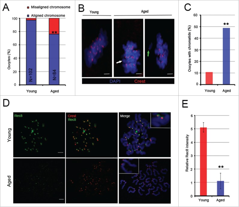
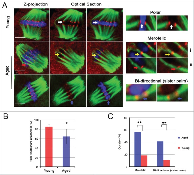
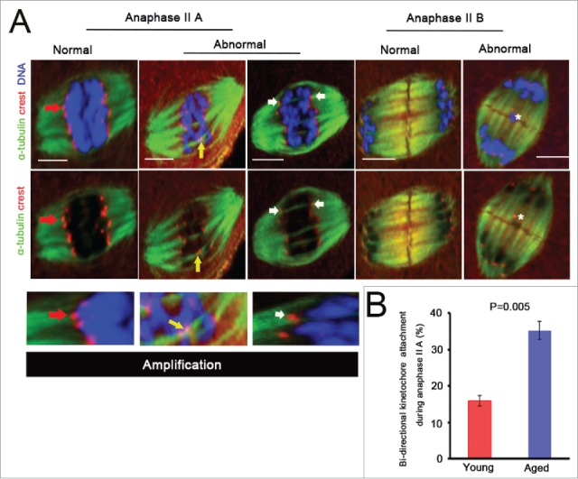
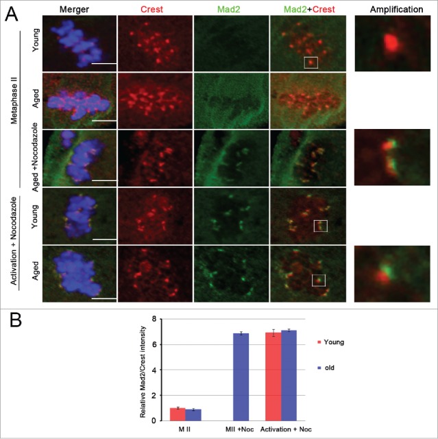
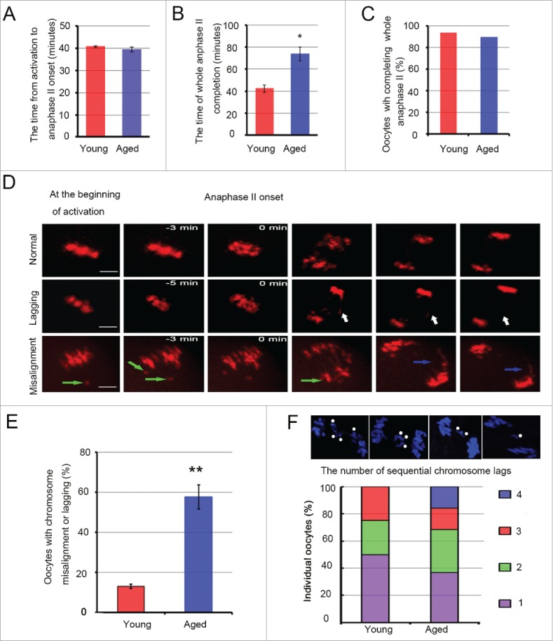
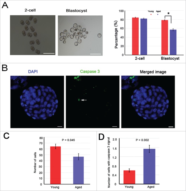
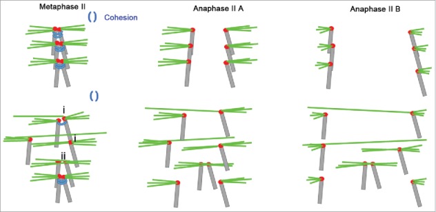
Mammalian oocyte chromosomes undergo 2 meiotic divisions to generate haploid gametes. The frequency of chromosome segregation errors during meiosis I increase with age. However, little attention has been paid to the question of how aging affects sister chromatid segregation during oocyte meiosis II. More importantly, how aneuploid metaphase II (MII) oocytes from aged mice evade the spindle assembly checkpoint (SAC) mechanism to complete later meiosis II to form aneuploid embryos remains unknown. Here, we report that MII oocytes from naturally aged mice exhibited substantial errors in chromosome arrangement and configuration compared with young MII oocytes. Interestingly, these errors in aged oocytes had no impact on anaphase II onset and completion as well as 2-cell formation after parthenogenetic activation. Further study found that merotelic kinetochore attachment occurred more frequently and could stabilize the kinetochore-microtubule interaction to ensure SAC inactivation and anaphase II onset in aged MII oocytes. This orientation could persist largely during anaphase II in aged oocytes, leading to severe chromosome lagging and trailing as well as delay of anaphase II completion. Therefore, merotelic kinetochore attachment in oocyte meiosis II exacerbates age-related genetic instability and is a key source of age-dependent embryo aneuploidy and dysplasia.

Keywords: aging; cohesion; kinetochore-microtubule attachment; meiosis II; oocytes; sister chromatids.


Figures

Figure 1.
Figure 1.
Aging causes centromeric cohesion deterioration, chromosome misalignment and premature sister chromatid segregation. (A) Percentages of young and aged MII oocytes with chromosomes aligned or misaligned at the spindle equator. Chromosomes were labeled with DAPI. In total, 84 aged and 102 young MII oocytes from more than 3 experiments were analyzed. (B) Localization of chromosomes and chromatids in MII oocytes from young and old mice. One chromatid aligned at the spindle equator is shown by a white arrow, and a green arrow indicates one misaligned chromatid. Red, centromere (Crest); blue, chromosome (DAPI). (C) The rate of MII oocytes with single chromatids in aged and young mice. The data were analyzed using a chi-square test. (D) Representative images of Rec8 protein immunostaining of aged and young MII oocytes. Green, Rec8; blue, chromosome (DAPI); red, centromere (Crest). (E) The relative Rec8 intensity of kinetochores was quantified. The data are the means ± SEM, ≥ 260 sister pairs of each group were analyzed. In A, C and E, **P <0.01. Scale bars in B and D, 5 μm.
Figure 2.
Figure 2.
Merotelic kinetochore and sister pair bi-directional attachments are increased significantly in aged MII oocytes. (A) Representative images of spindles and chromosomes in young and aged MII oocytes are shown. The right side shows the amplified images of polar kinetochore (white arrow), merotelic kinetochore (yellow and red arrow) and sister pair bi-directional (blue arrow) attachments. i shows 2 ends-on attachment, and ii shows lateral attachment. i and ii both belong to the merotelic kinetochore attachment category. DNA: blue, kinetochore: red, α-tubulin: green. Scale bar, 5 μm. (B) The rate of polar kinetochore attachment was analyzed in 14 young and 16 aged MII oocytes with 446 and 486 chromatids, respectively. The data represent the means ± SEM and were collected from 5 independent experiments, *P < 0.05. (C) The percentages of MII oocytes with merotelic kinetochore and sister pair bi-directional attachments in young and aged groups are shown. The data were analyzed using a chi-square test. **P < 0.01.
Figure 3.
Figure 3.
Bi-directional kinetochore attachment persists during oocyte anaphase II. (A) Different states of k-MT attachments and chromosomes during oocyte anaphase II are exhibited. Anaphase II is divided into anaphase II A and anaphase II B. In anaphase II A, kinetochores are attached by polar attachments (red arrow), Normal; some kinetochores are attached by merotelic attachments (white arrow), or one dyad is attached by sister pair bi-directional attachment (yellow arrow), Abnormal. In anaphase II B, no lagging chromosome, Normal; at least one lagging chromosome (white asterisk), Abnormal. Scale bar, 5μm. (B) Kinetochore attached by bipolar spindle microtubules in anaphase II A was quantified in 20 aged and 26 young oocytes. P = 0.005. The data were collected from 6 independent experiments and are shown as means ± SEM.
Figure 4.
Figure 4.
Aging has no impact on SAC mechanism. (A) Mad2 immunostaining was performed in young and aged MII oocytes and activated oocytes (60 min after Sr2+ addition) in the presence or absence of nocodazole. Images represent a single Z-plane. Scale bar, 5 μm. DNA: blue, kinetochore: red, Mad2: green. The right images are magnified pictures of the white framed areas. (B) Relative Mad2/Crest intensity was calculated in young and aged MII oocytes without (MII) or with nocodazole (MII + Noc) treatment and was calculated when nocodazole was added to the activation medium for 60 min (Activation + Noc). For each treatment, ≥ 310 kinetochores were analyzed. The data are the means ± SEM and were collected from 4 independent experiments.
Figure 5.
Figure 5.
Chromosome lagging and trailing, and the delay of anaphase II completion in aged oocytes. The whole anaphase II of 33 young and 31 aged oocytes from 9 independent experiments was observed by time-lapse imaging every 3 or 5 min. The chromosomes were labeled using Hoechst 3342 dye (blue changes into red in the figure). In A and B, the data represent the means ± SEM, *P = 0.05. In C, the data were analyzed using a chi-square test. (D) Sister chromatids localize at the metaphase plate and segregate into 2 separate chromosome structures in the second meiosis division, Normal. Single chromatids or sister pairs remain at the metaphase plate, or do not reach a spindle pole during anaphase II, Lagging (white arrow). Misaligned chromatid before anaphase onset (green arrow) and chromosomes have a severe trailing phenomenon (blue arrow), Misalignment. Scale bar, 10 μm. (E) The percentage of oocytes with chromosome misalignment or lagging was calculated in the young and aged groups. The data represent the means ± SEM, ** P < 0.01. (F) The number of lagging chromosomes was determined in 4 young and 19 aged, fixed oocytes during anaphase II; * represents one chromosome. The experiments were replicated more than 4 times.
Figure 6.
Figure 6.
Blastocyst quality in aged mice shows an obvious decrease. The 2-cell and blastocyst embryos were collected 48 and 96 h after in vitro culturing. In total, 121 young and 72 aged oocytes were activated. (A) Left: representative images of 2-cell and blastocyst embryos are shown, scale bar = 350 μm. Right: the percentages of 2-cell and blastocyst embryos, * P < 0.05. (B) The blastocysts were stained with cleaved caspase 3 (white arrow, green). DNA was counterstained with DAPI (blue). Scale bar, 10 μm. (C) The average cell number of one blastocyst. (D) The number of cells containing cleaved caspase 3 signal in one blastocyst. The data are the means ± SEM, and experiments were replicated more than 4 times.
Figure 7.
Figure 7.
Model for age-related sister chromatid segregation error in oocyte meiosis II. Aging causes the loss of centromere cohesion, which has a crucial role in stabilizing monopolar kinetochore attachment. Sister chromatids wrapped by centromeric cohesion are attached by amphitelic spindle microtubules for arrangement at the spindle equator and silencing the SAC mechanism in young oocytes. When centromeric cohesion deteriorates in the aged MII oocytes, monopolar kinetochore attachments may be replaced by (i) merotelic kinetochore or (ii) sister pair bi-directional attachments. These replacements generate new tensions that elude the SAC tension-monitoring mechanism. Some chromosomes attached by these replacement orientations show lagging and trailing because a single kinetochore is pulled simultaneously by 2 polar microtubules during anaphase II. Hence, bi-directional kinetochore attachment is the major reason for sister chromatid segregation errors during oocyte meiosis II in aged mice.

Similar articles

Cited by

References

    1. Jones KT, Lane SI. Molecular causes of aneuploidy in mammalian eggs. Development 2013; 140:3719-30; PMID:23981655; https://doi.org/10.1242/dev.090589 - DOI - PubMed
    1. Chiang T, Schultz RM, Lampson MA. Meiotic origins of maternal age-related aneuploidy. Biol Reprod 2012; 86:1-7; PMID:21957193; https://doi.org/10.1095/biolreprod.111.094367 - DOI - PMC - PubMed
    1. Holt JE, Lane SI, Jones KT. The control of meiotic maturation in mammalian oocytes. Curr Top Dev Biol 2013; 102:207-26; PMID:23287034 - PubMed
    1. Yun Y, Lane SI, Jones KT. Premature dyad separation in meiosis II is the major segregation error with maternal age in mouse oocytes. Development 2014; 141:199-208; PMID:24346700; https://doi.org/10.1242/dev.100206 - DOI - PMC - PubMed
    1. Salmon ED, Cimini D, Cameron LA, Deluca JG. Merotelic kinetochores in mammalian tissue cells. Philos Trans R Soc Lond B Biol Sci 2005; 360:553-68; PMID:15897180; https://doi.org/10.1098/rstb.2004.1610 - DOI - PMC - PubMed