. 2017 Sep;127(3):502-514.
doi: 10.1097/ALN.0000000000001719.

A Subregion of the Parabrachial Nucleus Partially Mediates Respiratory Rate Depression from Intravenous Remifentanil in Young and Adult Rabbits

Affiliations

A Subregion of the Parabrachial Nucleus Partially Mediates Respiratory Rate Depression from Intravenous Remifentanil in Young and Adult Rabbits

Justin R Miller et al. Anesthesiology. 2017 Sep.

Abstract

Background: The efficacy of opioid administration to reduce postoperative pain is limited by respiratory depression. We investigated whether clinically relevant opioid concentrations altered the respiratory pattern in the parabrachial nucleus, a pontine region contributing to respiratory pattern generation, and compared these effects with a medullary respiratory site, the pre-Bötzinger complex.

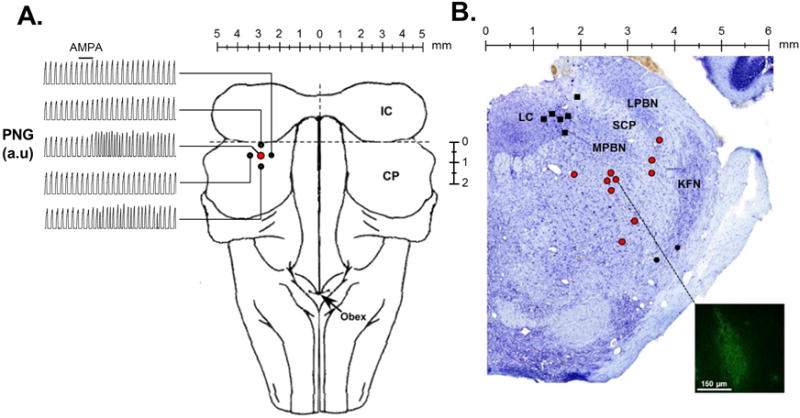
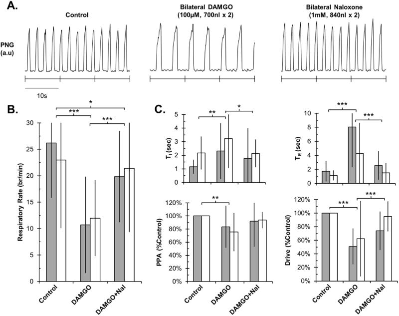
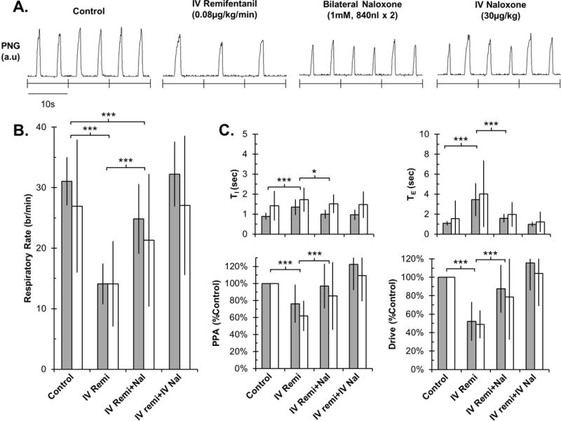
Methods: Studies were performed in 40 young and 55 adult artificially ventilated, decerebrate rabbits. We identified an area in the parabrachial nucleus where α-amino-3-hydroxy-5-methyl-4-isoxazolepropionic acid microinjections elicited tachypnea. Two protocols were performed in separate sets of animals. First, bilateral microinjections of the μ-opioid receptor agonist [D-Ala, N-MePhe, Gly-ol]-enkephalin (100 μM) into the "tachypneic area" determined the effect of maximal μ-opioid receptor activation. Second, respiratory rate was decreased with continuous IV infusions of remifentanil. The opioid antagonist naloxone (1 mM) was then microinjected bilaterally into the "tachypneic area" of the parabrachial nucleus to determine whether the respiratory rate depression could be locally reversed.

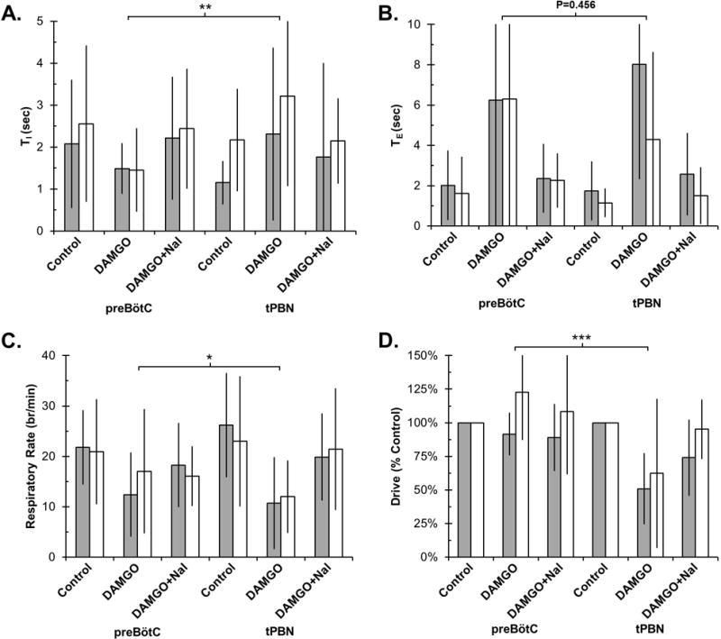
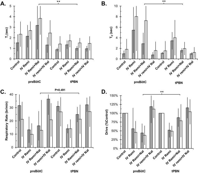
Results: Average respiratory rate was 27 ± 10 breaths/min. First, [D-Ala, N-MePhe, Gly-ol]-enkephalin injections decreased respiratory rate by 62 ± 20% in young and 45 ± 26% in adult rabbits (both P < 0.001). Second, during IV remifentanil infusion, bilateral naloxone injections into the "tachypneic area" of the parabrachial nucleus reversed respiratory rate depression from 55 ± 9% to 20 ± 14% in young and from 46 ± 20% to 18 ± 27% in adult rabbits (both P < 0.001). The effects of bilateral [D-Ala, N-MePhe, Gly-ol]-enkephalin injection and IV remifentanil on respiratory phase duration in the "tachypneic area" of the parabrachial nucleus was significantly different from the pre-Bötzinger complex.

Conclusions: The "tachypneic area" of the parabrachial nucleus is highly sensitive to μ-opioid receptor activation and mediates part of the respiratory rate depression by clinically relevant administration of opioids.


Conflict of interest statement

Disclosures

The authors state no conflicts of interest.

Figures

Figure 1
Figure 1
Injections of the glutamate receptor AMPA were used to functionally identify the “tachypneic area” in the parabrachial nucleus (tPBN). A: Dorsal brainstem image with representative records of the time-averaged phrenic neurogram (PNG, a.u.=arbitrary units). AMPA injection (solid bar) caused marked tachypnea in the tPBN (red circle), but little or no change in four surrounding sites 0.5 mm off-target (solid circles) in an individual adult rabbit. B: Location of fluorescent tracer or Chicago sky blue (700nl) injections into the LC (solid black squares), tPBN (red circles), and KFN (solid black circles) in Nissl stained tissue (Control Study). The inset depicts an example of the tracer spread which has been contrast enhanced to highlight the injection site. LC: locus coeruleus, LPBN: lateral parabrachial nucleus, MPBN: medial parabrachial nucleus, SCM: superior cerebellar peduncle, KFN: Kölliker Fuse nucleus.
Figure 2
Figure 2
Bilateral DAMGO injection into the tPBN significantly reduced respiratory rate and drive in young and adult rabbits, which was reversible with local naloxone. A: PNG tracing during control and after drug injections in an individual adult rabbit. B: Summary data for changes in respiratory rate. C: Summary data for changes in other respiratory parameters. Grey bars: young (n=14). White bars: adult (n=11). Mean ± SD. * P<0.05, ** P<0.01 and *** P<0.001 indicate significant differences between drug application conditions (linear mixed model with factors: drug application and developmental age). There were no differences in drug effects between young and adult animals.
Figure 3
Figure 3
Bilateral naloxone injection into the tPBN significantly reversed IV remifentanil-induced respiratory rate depression in young and adult rabbits. Residual changes from control were completely reversed with IV naloxone injections. A: PNG tracing during control conditions and after drug injections in an individual adult rabbit. B: Summary data for changes in respiratory rate. C: Summary data for changes in other respiratory parameters. Grey bars: young (n=12). White bars: adult (n=12). Mean±SD. * P<0.05; *** P<0.001 indicate significant differences between drug application conditions (linear mixed model, factors: drug application and developmental age). There were no differences in drug effects between young and adult animals.
Figure 4
Figure 4
Bilateral DAMGO injection into the tPBN had a significantly different effect on respiratory timing and drive compared to the preBötC. Values are displayed as Mean±SD for TI (A), TE (B), respiratory rate (C), and respiratory drive (D). Note differences in scale for TI and TE. Values on the left are from our previous study in the preBötC and values on the right are from our current tPBN study. PreBötC: Grey bars: young, n=8. White bars: adult, n=16. tPBN: Grey bars: young, n=14. White bars: adult, n=11. * P<0.05, ** P<0.01 and *** P<0.001 indicate significant differences in DAMGO effect between studies (linear mixed model, factors: study, drug application, developmental age).
Figure 5
Figure 5
Bilateral naloxone injection into the tPBN during IV remifentanil infusion had a significantly different effect on respiratory timing and drive compared to the preBötC. Values are displayed as Mean±SD for TI (A), TE (B), respiratory rate (C), and respiratory drive (D). Note differences in scale for TI and TE. Values on the left are from our previous study in which naloxone was injected into the preBötC and values on the right are from our current study. PreBötC: Grey bars: young, n=14. White bars: adult, n=16. tPBN: Grey bars: young, n=12. White bars: adult, n=12. ** P<0.01 indicate significant differences between study and drug application conditions (linear mixed model, factors: study, drug application, developmental age).
Figure 6
Figure 6
Hypothetical model of inputs determining (A) inspiratory and (B) expiratory phase duration. Intrinsic activity (Iintrinsic) is modified by opioid-sensitive inputs from the preBötzinger Complex (FpreBötC), the tachypneic area of the parabrachial nucleus (FtPBN) and additional brainstem sites (Fadditional). Systemic opioids reduce these inputs by the factor “r”. FpreBötC increases (+) inspiratory duration (TI) while FtPBN and Fadditional decrease (−) TI. Systemic opioids reduce these inputs resulting in a net increase in TI. All opioid-sensitive inputs decrease TE. Systemic opioids reduce these inputs resulting in a large increase in TE.

References

    1. Dahan A, Aarts L, Smith TW. Incidence, Reversal, and Prevention of Opioid-induced Respiratory Depression. Anesthesiology. 2010;112:226–38. - PubMed
    1. Hanna MH, Elliott KM, Fung M. Randomized, double-blind study of the analgesic efficacy of morphine-6-glucuronide versus morphine sulfate for postoperative pain in major surgery. Anesthesiology. 2005;102:815–21. - PubMed
    1. Lalley PM. Opiate slowing of feline respiratory rhythm and effects on putative medullary phase-regulating neurons. Am J Physiol Regul Integr Comp Physiol. 2006;290:R1387–96. - PubMed
    1. Mellen NM, Janczewski WA, Bocchiaro CM, Feldman JL. Opioid-induced quantal slowing reveals dual networks for respiratory rhythm generation. Neuron. 2003;37:821–6. - PMC - PubMed
    1. Dahan A, Romberg R, Teppema L, Sarton E, Bijl H, Olofsen E. Simultaneous measurement and integrated analysis of analgesia and respiration after an intravenous morphine infusion. Anesthesiology. 2004;101:1201–9. - PubMed