

CHARACTERISTICS OF EPIRETINAL MEMBRANE REMNANT EDGE BY OPTICAL COHERENCE TOMOGRAPHY AFTER PARS PLANA VITRECTOMY
- PMID: 28590966
- PMCID: PMC5505813
- DOI: 10.1097/IAE.0000000000001466
CHARACTERISTICS OF EPIRETINAL MEMBRANE REMNANT EDGE BY OPTICAL COHERENCE TOMOGRAPHY AFTER PARS PLANA VITRECTOMY
Abstract
Purpose: To evaluate the incidence, characteristics, and the progression of epiretinal membrane (ERM) remnant edge seen by optical coherence tomography after ERM peeling.
Methods: A retrospective chart review was conducted for 86 eyes of 85 consecutive patients who were diagnosed with ERM and underwent pars plana vitrectomy for epiretinal membrane peeling between 2013 and 2014. Data collected and analyzed included age, gender, preoperative and postoperative visual acuity, use of indocyanine green dye to stain internal limiting membrane, tamponade used after vitrectomy, ERM edge boundaries, presence of cystoid macular edema, and central foveal thickness.
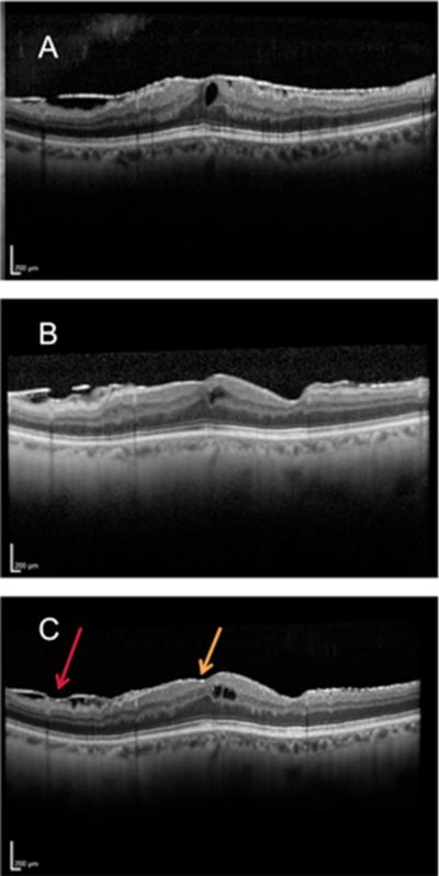
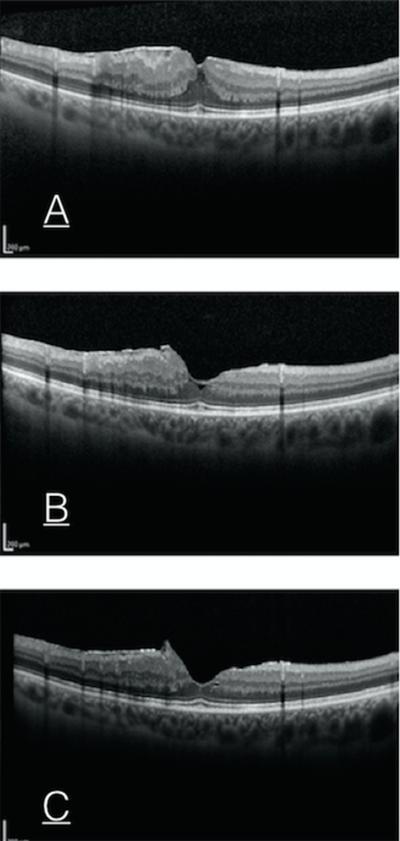
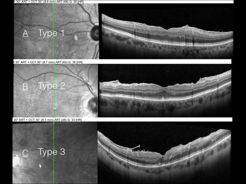
Results: An ERM remnant edge was detected in 33/86 study eyes (38.4%) at the first postoperative optical coherence tomography scan. Compared with those without an ERM remnant, patients with an ERM remnant after surgery were significantly older at baseline and had a higher incidence of ERM recurrence at their last visit. They were not significantly different in terms of gender, preoperative and postoperative visual acuity, reduction of central foveal thickness from baseline, proportion of eyes with preoperative ERM elevation on optical coherence tomography, presence of macular edema before surgery, intraoperative use of indocyanine green staining for ILM peeling, or tamponade used. Based on the edge morphology, we classified the ERM remnant into three types: Type 1 was flat and blended with the retina (14/33 eyes, 42.4%), Type 2 was flat but stepped (17/33 eyes, 51.5%), and Type 3 was elevated (2/33 eyes, 6.0%). A significantly higher risk of ERM recurrence was seen in Type 2 and Type 3 ERM remnants (75% and 100%, respectively) than Type 1 ERM remnants (10%).
Conclusion: An ERM remnant edge was detected by optical coherence tomography after ERM peeling in 38.4% of eyes. The presence of a postoperative ERM edge was associated with a higher risk of ERM recurrence, particularly in Type 2 and Type 3 ERM remnants.
Figures
References
-
- Mitchell P, Smith W, Chey T, et al. Prevalence and associations of epiretinal membranes. The Blue Mountains Eye Study, Australia. Ophthalmology. 1997;104:1033–1040. - PubMed
-
- You Q, Xu L, Jonas JB. Prevalence and associations of epiretinal membranes in adult Chinese: the Beijing eye study. Eye (Lond) 2008;22:874–879. - PubMed
-
- Gass JDM. Macular dysfunction caused by epiretinal membrane contraction. Stereoscopic Atlas of Macular Diseases: Diagnosis and Treatment. 1997:938–950.
-
- Park DW, Dugel PU, Garda J, et al. Macular pucker removal with and without internal limiting membrane peeling: pilot study. Ophthalmol. 2003;110:62–64. - PubMed
MeSH terms
Grants and funding
LinkOut - more resources
Full Text Sources
Other Literature Sources
Medical
Miscellaneous
