

Primary hepatic malignant fibrous histiocytoma combined with invasion of inferior vena cava: A case report and literature review
- PMID: 28591058
- PMCID: PMC5466236
- DOI: 10.1097/MD.0000000000007110
Primary hepatic malignant fibrous histiocytoma combined with invasion of inferior vena cava: A case report and literature review
Abstract
Rationale: Malignant fibrous histiocytoma (MFH), primary presented in liver, was very rare and displayed a poor prognosis because of high aggression. As a few of cases had been reported merely, we shared the case of primary hepatic MFH combined with invasion of inferior vena cava (IVC).
Patients concerns: A 69-year-old women presented with abdominal pain.
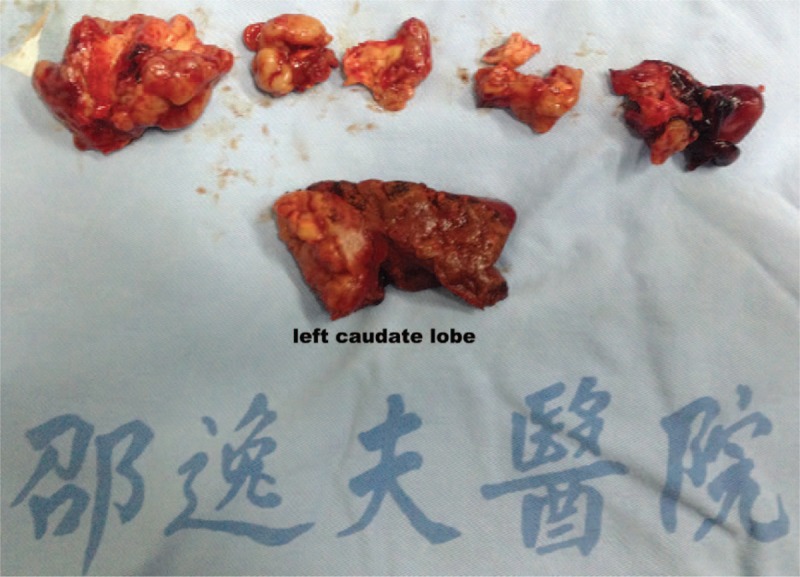
Diagnoses: Abdominal computed tomography and magnetic resonance imaging indicated a soft mass about 5.4 × 4.2 cm in the caudate lobe, accompanied with IVC invaded.
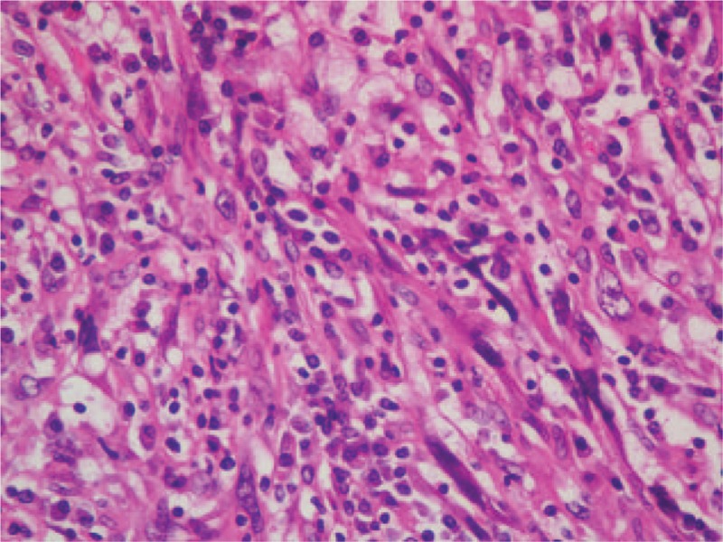
Interventions: After the multidisciplinary consultation, laparotomy was performed, followed by chemotherapy and radiotherapy. Primary hepatic MFH was demonstrated pathologically. Till now, the patient was alive for >22 months after surgery and no evidence of recurrence or distant metastasis was suspected.
Outcomes: We discussed the integrated procedure of diagnosis and treatment, combined with data from literature review.
Lessons: To our knowledge, the primary hepatic MFH combined with invasion of IVC was hardly reported. Despite the poor prognosis, the comprehensive treatment integrating the surgery, chemotherapy, and radiotherapy showed the satisfactory disease-free and overall survival. However, further investigations are definitely warranted.
Conflict of interest statement
The authors report no conflicts of interest.
Figures


Similar articles
-
Unusual Techniques for Preserving Surgical and Oncologic Safety in Hepatectomy of Advanced Adrenal Malignancy with Vena Cava and Liver Invasion.Ann Surg Oncol. 2018 Oct;25(11):3324-3325. doi: 10.1245/s10434-018-6657-5. Epub 2018 Jul 17. Ann Surg Oncol. 2018. PMID: 30019302
-
Primary hepatic malignant fibrous histiocytoma: a case report and review of the literature.Histol Histopathol. 2007 Dec;22(12):1337-42. doi: 10.14670/HH-22.1337. Histol Histopathol. 2007. PMID: 17701913
-
Primary malignant fibrous histiocytoma of the pancreas: benefit of the multidisciplinary approach.Eur J Gastroenterol Hepatol. 2010 Jun;22(6):765-8. Eur J Gastroenterol Hepatol. 2010. PMID: 20446353
-
Clinical characteristics of the primary hepatic malignant fibrous histiocytoma in China: case report and review of the literature.World J Surg Oncol. 2012 Jan 5;10:2. doi: 10.1186/1477-7819-10-2. World J Surg Oncol. 2012. PMID: 22221822 Free PMC article. Review.
-
Surgical challenges in the treatment of leiomyosarcoma of the inferior vena cava: analysis of two cases and brief review of the literature.Ann Vasc Surg. 2010 Aug;24(6):826.e13-7. doi: 10.1016/j.avsg.2010.02.039. Epub 2010 May 13. Ann Vasc Surg. 2010. PMID: 20471215 Review.
Cited by
-
Undifferentiated Pleomorphic Sarcoma of Liver: Case Report and Review of the Literature.Case Rep Pathol. 2018 Jul 17;2018:8031253. doi: 10.1155/2018/8031253. eCollection 2018. Case Rep Pathol. 2018. PMID: 30105111 Free PMC article.
References
-
- O’Brien JE, Stout AP. Malignant fibrous xanthomas. Cancer 1964;17:1445–55. - PubMed
-
- Weiss SW, Enzinger FM. Malignant fibrous histiocytoma: an analysis of 200 cases. Cancer 1978;41:2250–66. - PubMed
-
- Kransdorf MJ. Malignant soft-tissue tumors in a large referral population: distribution of diagnoses by age, sex, and location. Am J Roentgenol 1995;164:129–34. - PubMed
Publication types
MeSH terms
LinkOut - more resources
Full Text Sources
Other Literature Sources
Medical
Research Materials
