

A new technique for lipid core plaque detection by optical coherence tomography for prevention of peri-procedural myocardial infarction: A case report
- PMID: 28591063
- PMCID: PMC5466241
- DOI: 10.1097/MD.0000000000007125
A new technique for lipid core plaque detection by optical coherence tomography for prevention of peri-procedural myocardial infarction: A case report
Abstract
Rationale: Percutaneous coronary intervention (PCI) provides effective revascularization of atherosclerotic coronary arteries but the invasive nature of treatment can result in complications.
Patient concerns: A 53-year old man underwent coronary angiography due to chest pain with minimal ST-segment elevation in the inferior leads of the electrocardiogram.
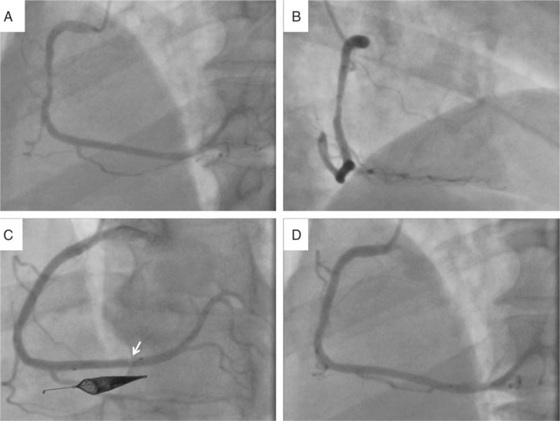
Diagnosis: We proceeded directly to coronary angiography and delineated a moderate stenosis with haziness in the mid right coronary artery (RCA).
Interventions: Expert analysis of the pre-intervention OCT imaging demonstrated a large lipid core plaque (LCP), upstream of the culprit site, with minimal thrombus burden. Subsequent implantation of a bioresorbable vascular scaffold, protected with distal deployment of a filter protection device provided an excellent result with retrieval of plaque material. Post-hoc attenuation analysis confirmed the presence of large LCP.
Outcomes: A post-procedural transthoracic echocardiogram confirmed good left ventricular function with no regional wall motion abnormality. An excellent clinical outcome was achieved.
Lessons: Optical coherence tomography (OCT) derived attenuation analysis can provide with qualitative and quantitative detailed evaluation of the underlying plaque substrate. Our case shows OCT can provide the interventionist with qualitative and qualitative assessment of large LCP for prevention of periprocedural complications, which may improve outcome for PCI.
Conflict of interest statement
Conflicts of interest: Erasmus MC has a patent licensing agreement with Terumo Corporation in the area of OCT imaging. Dr Van Soest has the right to receive royalties as part of this agreement. Dr Johnson has received speaker and consultancy fees from St Jude Medical and Terumo Corporation. The other authors report no relationships that could be construed as a conflict of interest.
Figures

Similar articles
-
Optical coherence tomography guided successful treatment without stent implantation in a patient with non-ST-segment elevation myocardial infarction caused by plaque rapture: A case report.Medicine (Baltimore). 2018 Dec;97(50):e13669. doi: 10.1097/MD.0000000000013669. Medicine (Baltimore). 2018. PMID: 30558071 Free PMC article.
-
Predictors of periprocedural (type IVa) myocardial infarction, as assessed by frequency-domain optical coherence tomography.Circ Cardiovasc Interv. 2012 Feb 1;5(1):89-96, S1-6. doi: 10.1161/CIRCINTERVENTIONS.111.965624. Epub 2012 Jan 31. Circ Cardiovasc Interv. 2012. PMID: 22298799
-
[Impact of inflammatory reaction levels and culprit plaque characteristics on preprocedural thrombolysis in myocardial infarction flow grade in patients with ST-segment elevation myocardial infarction].Zhonghua Xin Xue Guan Bing Za Zhi. 2021 Feb 24;49(2):150-157. doi: 10.3760/cma.j.cn112148-20200531-00452. Zhonghua Xin Xue Guan Bing Za Zhi. 2021. PMID: 33611901 Chinese.
-
Optical coherence tomography-guided primary percutaneous coronary intervention in ST-segment elevation myocardial infarction patients: a pilot study.Can J Cardiol. 2014 Apr;30(4):420-7. doi: 10.1016/j.cjca.2013.12.016. Epub 2013 Dec 30. Can J Cardiol. 2014. PMID: 24680171
-
In vivo imaging of complicated atherosclerotic plaque - role of optical coherence tomography (OCT).Rom J Morphol Embryol. 2018;59(2):469-478. Rom J Morphol Embryol. 2018. PMID: 30173250 Review.
References
-
- van Soest G, Regar E, Goderie TP, et al. Pitfalls in plaque characterization by OCT: image artifacts in native coronary arteries. JACC Cardiovasc Imaging 2011;4:810–3. - PubMed
-
- Manfrini O, Mont E, Leone O, et al. Sources of error and interpretation of plaque morphology by optical coherence tomography. Am J Cardiol 2006;98:156–9. - PubMed
-
- Herrmann J. Peri-procedural myocardial injury: 2005 update. Eur Heart J 2005;26:2493–519. - PubMed
-
- Porto I, Selvanayagam JB, Van Gaal WJ, et al. Plaque volume and occurrence and location of periprocedural myocardial necrosis after percutaneous coronary intervention: Insights from delayed-enhancement magnetic resonance imaging, thrombolysis in myocardial infarction myocardial perfusion grade analysis, and intravascular ultrasound. Circulation 2006;114:662–9. - PubMed
-
- Prasad A, Herrmann J. Myocardial infarction due to percutaneous coronary intervention. N Engl J Med 2011;364:453–64. - PubMed
Publication types
MeSH terms
LinkOut - more resources
Full Text Sources
Other Literature Sources
Medical
Miscellaneous
