. 2017 Jun 7;12(6):e0178969.
doi: 10.1371/journal.pone.0178969. eCollection 2017.

AKT and its related molecular feature in aged mice skin

Affiliations

AKT and its related molecular feature in aged mice skin

Haiyan Chen et al. PLoS One. .

Abstract

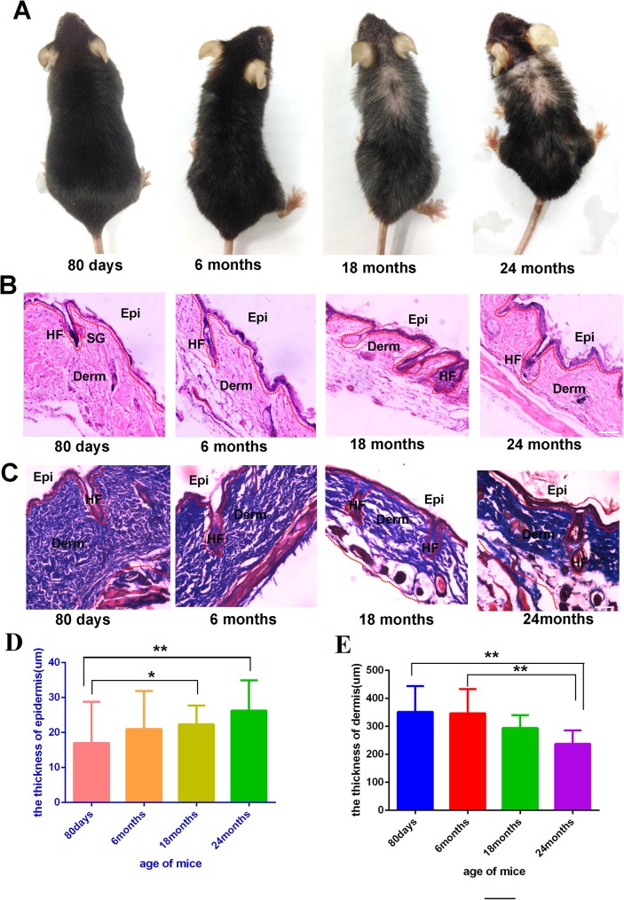
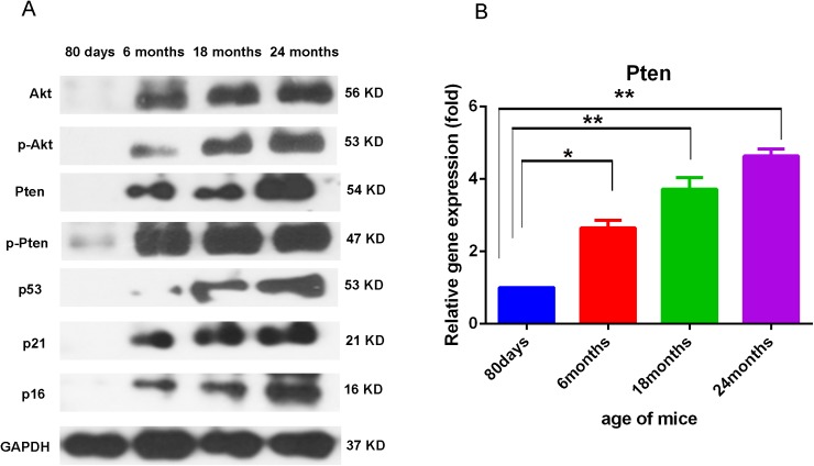
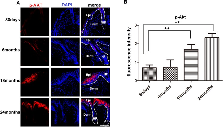
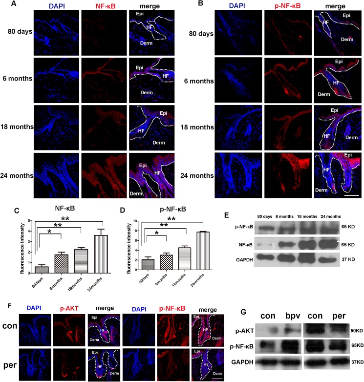
Previous studies suggest that Akt signaling promotes tissue regeneration and decreased Akt activities are found in aged tissues. However, this study finds that the expression and activation levels of Akt in the mice skin increased with age. Additionally, the expression levels of Pten, p16, p21 and p53 also elevated with increased age. Immuno-fluorescence analysis showed that Akt phosphorylation found in the epidermal cells (with increased levels of NF-κB activation) were also found. In vivo inhibition of AKT activity result in reduced NF-κB activation. Our results suggest that increasing Akt/ NF-κB is a crucial mediator of skin aging, which can increase the susceptibility of cell transformation.


Conflict of interest statement

Competing Interests: The authors have declared that no competing interests exist.

Figures

Fig 1
Fig 1
Representative images of mice of different ages, 80 days mice (n = 6), 6 months mice (n = 6), 18 months mice (n = 6), 24 months mice (n = 3) (A). HE staining of the skin from different ages mice (B,C), For HE and Masson staining, representative images from 8–16 tissue sections in 2–6 mice are shown the thickness of full skin and epidermis of different age mice(D,E).Data are expressed as the mean ± s.e.m. *** P<0.005, unpaired t-test, two-tailed. Scale bars, 50 μm.
Fig 2
Fig 2. Akt activation levels in the skin of mice in different ages.
Representative results of Western blotting analysis of skin tissues from mice aged 80 days, 6 months,18 months and 24 months for the levels of Pten, p-Pten, Akt, p-Akt (Ser 473), p53, p21 and p16(A). For all Western blot analysis, data are representative of 3–5 independent experiments. Real time PCR analysis of skins from different age mice showed higher PTEN expression with the mice age grows (B). For all real-time PCR analyses, gene expression was normalized to GAPDH with 40 cycles, data are represented as the mean ± SD, and N = 3.
Fig 3
Fig 3. Levels of p-Akt in the skin of mice in different ages.
(A)(IF?)Immuno-fluorescence analysis barely detected the presence of p-Akt positive cells in the skin of 80 days and 6 months old mice, but detected evident p-Akt positive cells in the epidermis and hair follicle of 18 and 24 months old mice. For all IF analyses, representative images from 8–16 tissue sections in 3–6 mice are shown. (B) Fluorescence intensity of p-Akt of 80days, 6months, 18 months, 24months old mice were measured by image J. Scale bars, 20 μm for microscopic images. Data are expressed as the mean ± s.e.m. *** P<0.005, unpaired t-test, two-tailed.
Fig 4
Fig 4. Levels of NF-κB, p-NF-κB in the skin of mice in different ages.
(A, B) Immuno-fluorescence analysis showed increasing levels of NF-κB and p-NF-κB in the skin of mice with aging. Scale bars, 20 μm. (C,D) Fluorescence intensity of NF-κB, p-NF-κB of 80days,6months,18months, 24months old mice were measured by imageJ. (E) Western blotting analysis showed the levels of NF-κB and p-NF-κB in the skin tissue of mice aged 80 days, 6 months, 18 months and 24 months. (F) Immuno-fluorescence analysis showed increasing levels of p-AKT and p-NF-κB in the skin of 12 months mice treated with perifosine and the littermate without perifosine treated (N = 3). Scale bars, 20 μm. (G) Western blotting analysis showed the levels of p-AKT and p-NF-κB in the skin of 12 months mice treated with perifosine and 80 days mice which treated with the bpv(phen), the littermate no-treated mice as control group (N = 3).

References

    1. Ressler S, Bartkova J, Niederegger H, Bartek J, Scharffetter-Kochanek K, Jansen-Durr P, et al. p16INK4A is a robust in vivo biomarker of cellular aging in human skin. Aging cell. 2006;5(5):379–89. doi: 10.1111/j.1474-9726.2006.00231.x - DOI - PubMed
    1. Blanco-Aparicio C, Renner O, Leal JF, Carnero A. PTEN, more than the AKT pathway. Carcinogenesis. 2007;28(7):1379–86. doi: 10.1093/carcin/bgm052 - DOI - PubMed
    1. Xia X, Park E, Liu B, Willette-Brown J, Gong W, Wang J, et al. Reduction of IKKalpha expression promotes chronic ultraviolet B exposure-induced skin inflammation and carcinogenesis. The American journal of pathology. 2010;176(5):2500–8. PubMed Central PMCID: PMC2861114. doi: 10.2353/ajpath.2010.091041 - DOI - PMC - PubMed
    1. Tatar M, Kopelman A, Epstein D, Tu MP, Yin CM, Garofalo RS. A mutant Drosophila insulin receptor homolog that extends life-span and impairs neuroendocrine function. Science. 2001;292(5514):107–10. doi: 10.1126/science.1057987 - DOI - PubMed
    1. Datta SR, Brunet A, Greenberg ME. Cellular survival: a play in three Akts. Genes & development. 1999;13(22):2905–27. - PubMed