. 2017 Jun 6;112(11):2327-2335.
doi: 10.1016/j.bpj.2017.04.041.

Blocking of Single α-Hemolysin Pore by Rhodamine Derivatives

Affiliations

Blocking of Single α-Hemolysin Pore by Rhodamine Derivatives

Tatyana I Rokitskaya et al. Biophys J. .

Abstract

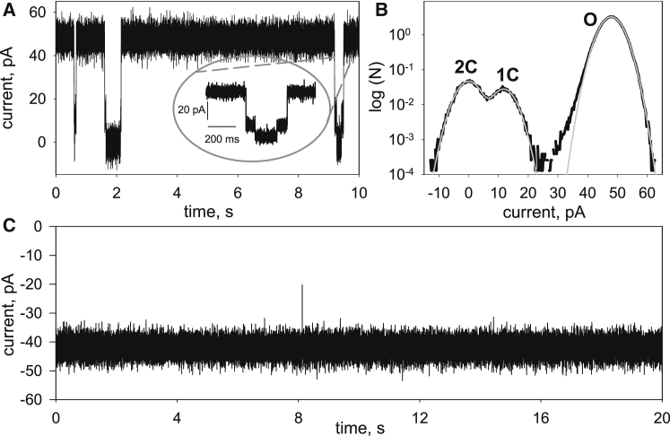
Measurements of ion conductance through α-hemolysin pore in a bilayer lipid membrane revealed blocking of the ion channel by a series of rhodamine 19 and rhodamine B esters. The longest dwell closed time of the blocking was observed with rhodamine 19 butyl ester (C4R1), whereas the octyl ester (C8R1) was of poor effect. Voltage asymmetry in the binding kinetics indicated that rhodamine derivatives bound to the stem part of the aqueous pore lumen. The binding frequency was proportional to a quadratic function of rhodamine concentrations, thereby showing that the dominant binding species were rhodamine dimers. Two levels of the pore conductance and two dwell closed times of the pore were found. The dwell closed times lengthened as the voltage increased, suggesting impermeability of the channel for the ligands. Molecular docking analysis revealed two distinct binding sites within the lumen of the stem of the α-hemolysin pore for the C4R1 dimer, but only one binding site for the C8R1 dimer. The blocking of the α-hemolysin nanopore by rhodamines could be utilized in DNA sequencing as additional optical sensing owing to bright fluorescence of rhodamines if used for DNA labeling.


Figures

Figure 1
Figure 1
Shown here are the traces of current through the single αHL channel without rhodamines (A) or in the presence of 20 μM C2R1 (B), 20 μM C4R1 (C), 15 μM C8R1 (D), or 20 μM C12R1 (E). The inset to (B) demonstrates in more detail two blocking states of the channel (1C and 2C). The applied potential was +50 mV.
Figure 2
Figure 2
(A) Shown here are the traces of the current through the single αHL channel in the presence of 5 μM C4R1 at the applied potential +50 mV (A) or −50 mV (C). (B) Representative current amplitude histogram of the recording at +50 mV was fitted by a sum of three Gaussians (gray curves). Positions of the peaks and widths of distributions are 0 ± 3.4 pA (marked 2C), 11.8 ± 3.3 pA (marked 1C) and 47.9 ± 3.3 pA (marked O).
Figure 3
Figure 3
(A) Shown here is the log-binned dwell open time histogram of the single αHL pore with approximation by a single exponent in the presence of 25 μM C2R1 (τon = 0.77 s). The applied potential was +50 mV. (B). Shown here is the inverse mean dwell open time (1/τon) of the αHL channel as a function of C2R1 concentration. The line represents approximation of the data by the power function [C2R1]n with n = 2.2.
Figure 4
Figure 4
(A) Shown here are the log-binned dwell blockage time histograms for C2R1 and C4R1 binding with the single αHL channel. Distributions of blockage times were analyzed by logarithmic double exponential fitting (τoff1 = 12 ms and τoff2 = 0.18 s for C2R1, medium dashed line; τoff1 = 18 ms and τoff2 = 0.88 s for C4R1, solid line). (B) Shown here is a trace of the current through the single αHL pore in the presence of 10 μM C4R1, demonstrating, in detail, two blocked states of the pore designating two types of events corresponding to τoff1 and τoff2. (C) Shown here is the probability of the single αHL channel being in unblocked state versus applied potential in the presence of 10 μM C4R1. (D) Shown here is the dependence of τon (solid circles), τoff1 (dotted circles), and τoff2 (open circles) on BLM voltage in the presence of 10 μM C4R1.
Figure 5
Figure 5
Shown here are the traces of the current through the single αHL channel in the presence of 15 μM C2RB at applied potential +50 mV (A) or −50 mV (C). The inset to (A) demonstrates in more detail the blocking states of the channel. (B) Shown here are the log-binned dwell blockage time histograms for C2RB and C4RB binding with the single αHL channel. Distributions of blockage times were analyzed by logarithmic double exponential fitting (τoff1 = 6 ms and τoff2 = 24 s for C2RB, medium dashed line; τoff1 = 15 ms and τoff2 = 72 ms for C4RB, solid line).
Figure 6
Figure 6
Shown here are the locations of rhodamine derivative binding sites in the αHL pore down the membrane center (A, side view; B, bottom view). The dimer of the rhodamine derivatives is marked in blue for C2R1, green for C4R1, and red for C8R1. To see this figure in color, go online.
Figure 7
Figure 7
(A) Shown here is the density of location of the bridging oxygen atoms of the rhodamines dimer in the αHL pore. Blue and yellow colors mean higher and lower probability, respectively. (B) Shown here are the histograms of the distribution of the docking occurrence in the αHL pore on the distance from the membrane center. The borders of the lipid bilayer are marked by black dashed lines. The dimer of the rhodamine derivatives is marked in blue for C2R1, green for C4R1, and red for C8R1. To see this figure in color, go online

References

    1. Menestrina G. Ionic channels formed by Staphylococcus aureus α-toxin: voltage-dependent inhibition by divalent and trivalent cations. J. Membr. Biol. 1986;90:177–190. - PubMed
    1. Krasilnikov O.V., Sabirov R.Z. Ion transport through channels formed in lipid bilayers by Staphylococcus aureus α-toxin. Gen. Physiol. Biophys. 1989;8:213–222. - PubMed
    1. Song L., Hobaugh M.R., Gouaux J.E. Structure of staphylococcal α-hemolysin, a heptameric transmembrane pore. Science. 1996;274:1859–1866. - PubMed
    1. Gouaux J.E., Braha O., Bayley H. Subunit stoichiometry of staphylococcal α-hemolysin in crystals and on membranes: a heptameric transmembrane pore. Proc. Natl. Acad. Sci. USA. 1994;91:12828–12831. - PMC - PubMed
    1. Bayley H., Cremer P.S. Stochastic sensors inspired by biology. Nature. 2001;413:226–230. - PubMed

LinkOut - more resources