. 2017 Jul 25;8(30):49807-49823.
doi: 10.18632/oncotarget.17971.

Maintenance of cancer stemness by miR-196b-5p contributes to chemoresistance of colorectal cancer cells via activating STAT3 signaling pathway

Affiliations

Maintenance of cancer stemness by miR-196b-5p contributes to chemoresistance of colorectal cancer cells via activating STAT3 signaling pathway

Dong Ren et al. Oncotarget. .

Abstract

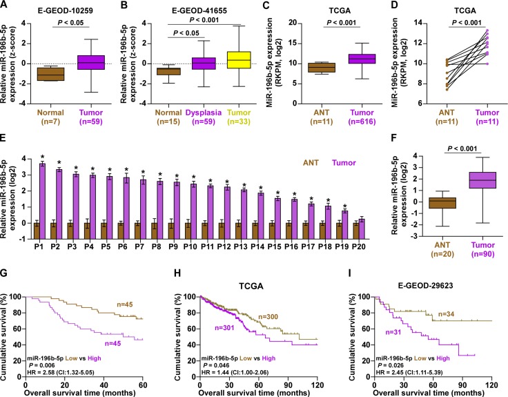
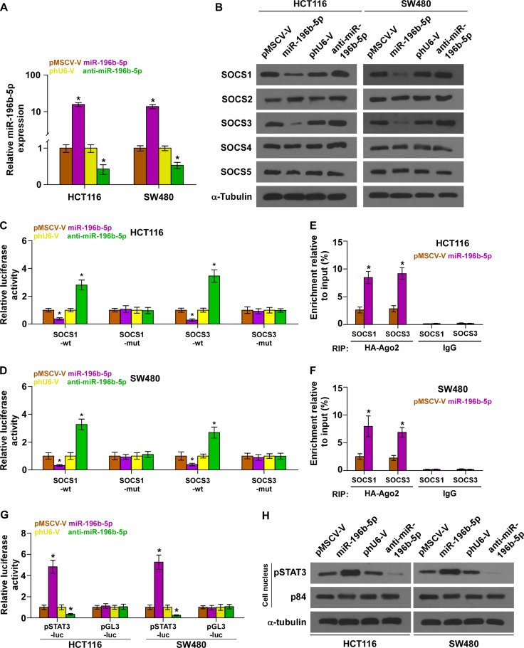
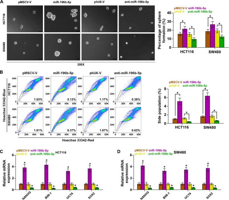
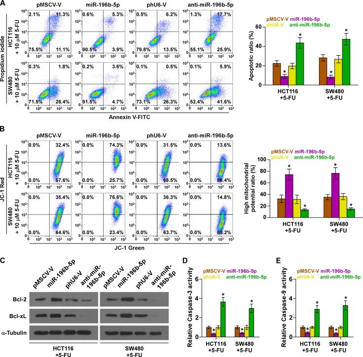
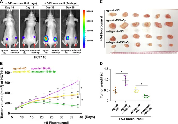
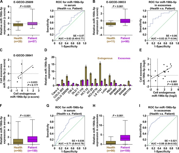
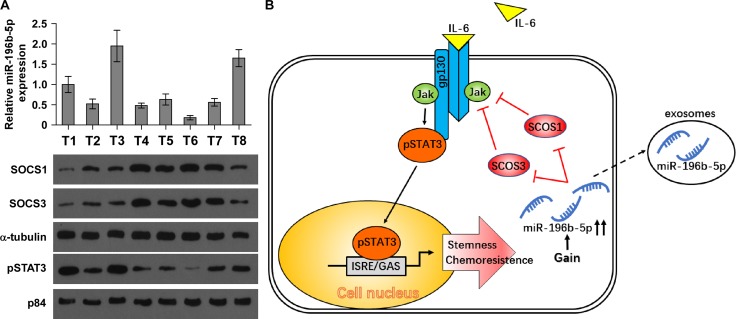
Emerging studies indicated that cancer stem cells represent a subpopulation of cells within the tumor that is responsible for chemotherapeutic resistance. However, the underlying mechanism is still not clarified yet. Here we report that miR-196b-5p is dramatically upregulated in CRC tissues and high expression of miR-196b-5p correlates with poor survival in CRC patients. Moreover, recurrent gains (amplification) contribute to the miR-196b-5p overexpression in CRC tissues. Silencing miR-196b-5p suppresses spheroids formation ability, the fraction of SP cells, expression of stem cell factors and the mitochondrial potential, and enhances the apoptosis induced by 5-fluorouracil in CRC cells; while ectopic expression of miR-196b-5p yields an opposite effect. In addition, downregulation of miR-196b-5p resensitizes CRC cells to 5-fluorouracil in vivo. Our results further demonstrate that miR-196b-5p promotes stemness and chemoresistance of CRC cells to 5-fluorouracil via targeting negative regulators SOCS1 and SOCS3 of STAT3 signaling pathway, giving rise to activation of STAT3 signaling. Interestingly, miR-196b-5p is highly enriched in the serum exosomes of patients with CRC compared to the healthy control subjects. Thus, our results unravel a novel mechanism of miR-196b-5p implicating in the maintenance of stem cell property and chemotherapeutic resistance in CRC, offering a potential rational registry of anti-miR-196b-5p combining with conventional chemotherapy against CRC.

Keywords: CRC; STAT3 signaling pathway; cancer stem cell; chemotherapeutic resistance; miR-196b-5p.


Conflict of interest statement

CONFLICTS OF INTEREST

No conflicts of interest were declared.

Figures

Figure 1
Figure 1. miR-196b-5p is upregulated in CRC and correlated with poor prognosis
(AC) miR-196b-5p expression levels was markedly upregulated in CRC tissues as assessed by analyzing the E-GEOD-10259, E-GEOD-41655 and TCGA of CRC miRNA sequencing datasets. (D) Real-time PCR analysis of miR-196b-5p in 11 primary CRC tissues compared with the matched adjacent normal tissues (ANT). (E) Real-time PCR analysis of miR-196b-5p expression in 20 paired collected CRC tissue samples. Transcript levels were normalized to U6 expression. Each bar represents the mean values ± SD of three independent experiments. *P < 0.05. (F) miR-196b-5p expression levels was markedly upregulated in CRC tissues compared with the matched adjacent normal tissues (ANT). (ANT, n = 20; CRC, n = 90). P < 0.001. (G) Kaplan–Meier analysis of overall survival curves of patients with CRC with high miR-196b-5p expression (> median, n = 45) versus low miR-196b-5p expression (< median, n = 45). P < 0.001, log-rank test. (H and I) Kaplan–Meier analysis of overall survival curves of CRC patients datasets from TCGA and E-GEOD-29623.
Figure 2
Figure 2. miR-196b-5p activates STAT3 signaling via targeting multiple negative regulators of STAT3 signaling
(A) Real-time PCR analysis of miR-196b-5p expression in the indicated cells. Transcript levels were normalized by U6 expression. Error bars represent the mean ± SD of three independent experiments. *P < 0.05. (B) Western blotting of SOCS1, SOCS2, SOCS3, SOCS4 and SOCS5 expression in the indicated cells. α-Tubulin served as the loading control. (C and D) Luciferase assay of cells transfected with pmirGLO-3′UTR reporter of SOCS1 and SOCS3 in miR-196b-5p overexpressing and silencing HCT116 and SW480 cells, respectively. Error bars represent the mean ± SD of three independent experiments. *P < 0.05. (E and F) MiRNP IP assay showing the association between miR-196b-5p and SOCS1,SOCS3 transcripts in HCT116 and SW480 cells. Pulldown of IgGantibody served as the negative control. Error bars represent the mean ± SD of three independent experiments. *P < 0.05. (G) STAT3 transcriptional activity was assessed by luciferase reporter constructs in the indicated cells. Error bars represent the mean ± SD of three independent experiments. *P < 0.05. (H) Western blotting of nuclear STAT3 expression. The nuclear protein p84 was used as the nuclear protein marker.
Figure 3
Figure 3. miR-196b-5p promotes stem cell properties in CRC cells
(A) Representative images of spheroids formed at 200-fold magnification were counted. Histograms showed the mean number of spheroids formed. Scale bars, 50 mm. Error bars represent the mean ± SD of three independent experiments. *P < 0.05. (B) Hoechst 33342 dye exclusion assay showed that overexpressing miR-196b-5p promoted the fraction of side population, whereas silencing miR-196b-5p decreased the fraction. Error bars represent the mean ± SD of three independent experiments. *P < 0.05. (C and D) Real-time PCR analysis of OCT4A, SOX2, NANOG and BMI-1 expression in the indicated cells. GAPDH was used as the loading control. Error bars represent the mean ± SD of three independent experiments. *P < 0.05.
Figure 4
Figure 4. miR-196b-5p promotes chemoresistance in CRC cells in vitro
(A) Annexin V-FITC/PI staining of the indicated cells under treatment of 5-FU. Error bars represent the mean ± SD of three independent experiments. *P < 0.05. (B) The JC-1 staining the indicated cells under treatment of 5-FU. Error bars represent the mean ± SD of three independent experiments. *P < 0.05. (C) Western blotting analysis of Bcl-2 and Bcl-xL in the indicated cells under treatment of 5-FU. (D and E) Analysis of the activities of caspase-3 (D) and caspase-9 (E) were detected by the cleaved forms of these two proteins. Error bars represent the mean ± SD of three independent experiments. *P < 0.05.
Figure 5
Figure 5. Inhibition of miR-196b-5p sensitizes CRC cells to 5-FU in vivo
(A) Representative images of tumor-bearing mice. The representative images were captured on day 14 and day 38 after inoculating HCT116 cells respectively. The intraperitoneal injection of 5-FU (50 mg/kg.d) started from two weeks after inoculating HCT116 cells and lasted for 24 days, namely 38 day after inoculating HCT116 cells. (B) Tumor volumes in the miR-196b-5p–overexpressing, miR-196b-5p–silenced, and control groups were measured on the indicated days. Data presented are the mean ± SD (C) Mice were euthanized, and tumors from each experimental group were excised. (D) Tumor weights of each group.
Figure 6
Figure 6. miR-196b-5p is detected in the serum and serum exosomes of CRC patients
(A) miR-196b-5p expression was elevated in the serum of CRC patients compared with the healthy controls from E-GEOD-25609 dataset (left panel) and ROC curve for miR-196b-5p in the serum of the healthy and CRC patients (right panel). (B) miR-196b-5p expression was elevated in the serum exosomes of CRC patients compared with the healthy controls from E-GEOD-39833 dataset (left panel) and ROC curve for miR-196b-5p in the serum exosomes of the healthy and CRC patients (right panel). (C) Correlation between mRNA expression levels of miR-196b-5p in 6 different CRC cells and the concentration of miR-196b-5p in their respective exosomes. (D) Endogenous expression of miR-196b-5p in diffetent CRC cell lines and miR-196b-5p expression in the exosomes from the supernatant of diffetent CRC cell lines. (E) Correlation between mRNA expression levels of miR-196b-5p in different CRC cells and the concentration of miR-196b-5p in their respective exosomes. (F) miR-196b-5p expression was elevated in the serum of CRC patients compared with the healthy controls (Health, n = 90; CRC, n = 150). P < 0.001. (G) ROC curve for miR-196b-5p in the serum of the healthy and CRC patients. (H) miR-196b-5p expression was elevated in the serum exosomes of CRC patients compared with the healthy controls (Health, n = 90; CRC, n = 150). P < 0.001. (I) ROC curve for miR-196b-5p in the serum exosomes of the healthy and CRC patients.
Figure 7
Figure 7. Clinical relevance of miR-196b-5p with SOCS1, SOCS3 and STAT3 signaling activity in human CRC tissues
(A) Analysis of miR-196b-5p expression with protein expression of SOCS1, SOCS3 and nuclear pSTAT3 in 8 CRC tissues. U6 was used as the control for RNA loading. miR-196b-5p expression levels were normalized to that miR-196b-5p expression of sample one. Loading controls were α-tubulin and p84 for the cytoplasmic and nuclear fractions. Each bar represents the mean ± SD of three independent experiments. *P < 0.05. (B) Hypothetical model illustrating that constitutive activation of the STAT3 signaling pathway by miR-196b-5p epigenetic disruption of multiple negative feedback loops contributes to the maintenance of stemness and chemoresistance in CRC cells.

Similar articles

Cited by

References

    1. Jemal A, Siegel R, Ward E, Hao Y, Xu J, Thun MJ. Cancer statistics, 2009. CA Cancer J Clin. 2009;59:225–249. - PubMed
    1. Ricci-Vitiani L, Fabrizi E, Palio E, De Maria R. Colon cancer stem cells. J Mol Med (Berl) 2009;87:1097–1104. - PubMed
    1. Thota R, Pauff JM, Berlin JD. Treatment of metastatic pancreatic adenocarcinoma: a review. Oncology (Williston Park) 2014;28:70–74. - PubMed
    1. Singh SK, Hawkins C, Clarke ID, Squire JA, Bayani J, Hide T, Henkelman RM, Cusimano MD, Dirks PB. Identification of human brain tumour initiating cells. Nature. 2004;432:396–401. - PubMed
    1. Park EK, Lee JC, Park JW, Bang SY, Yi SA, Kim BK, Park JH, Kwon SH, You JS, Nam SW, Cho EJ, Han JW. Transcriptional repression of cancer stem cell marker CD133 by tumor suppressor p53. Cell Death Dis. 2015;6:e1964. - PMC - PubMed

MeSH terms

Substances