

Biological Evaluation of a Fluorescent-Imaging Agent for Medullary Thyroid Cancer in an Orthotopic Model
- PMID: 28591772
- PMCID: PMC5587064
- DOI: 10.1210/jc.2017-00573
Biological Evaluation of a Fluorescent-Imaging Agent for Medullary Thyroid Cancer in an Orthotopic Model
Abstract
Context: The primary and definitive treatment of medullary thyroid cancer (MTC) is surgical resection. Recurrent or residual disease is typically a result of incomplete surgical removal.
Objective: Our objective is to develop a compound that assists in intraoperative visualization of cancer, which would have the potential to improve surgical cure rates and outcomes.
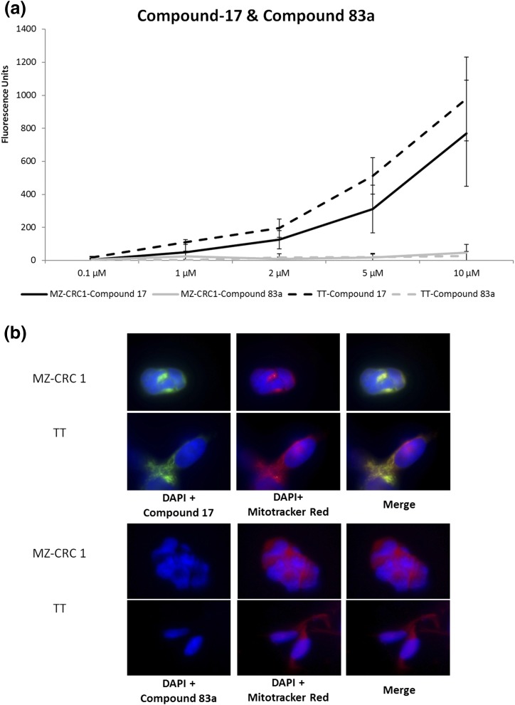
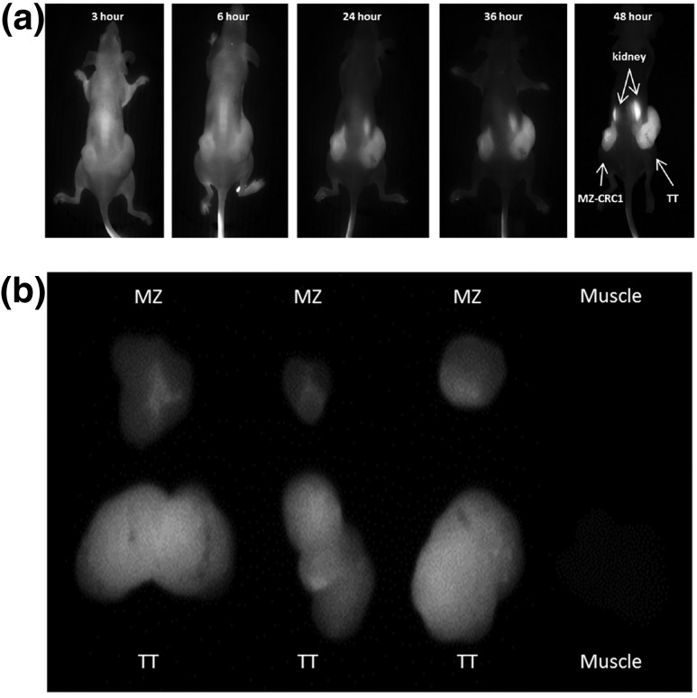
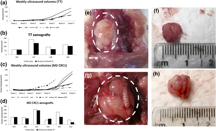
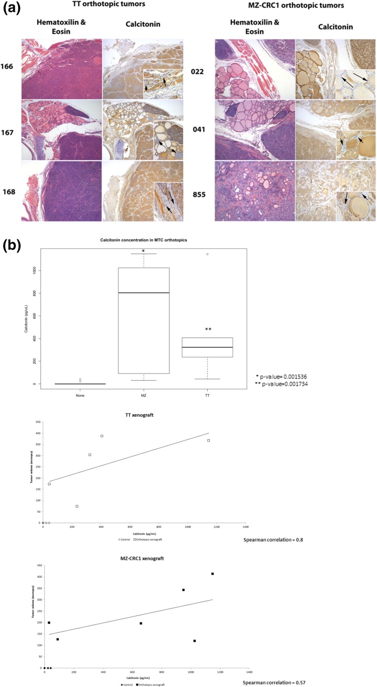
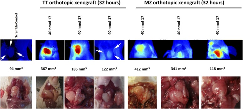
Results: We report the biological characterization of Compound-17, which is labeled with IRdye800, allowing fluorescent visualization of MTC mouse models. We found that the agent has high affinity for two human MTC cell lines (TT and MZ-CRC1) in vitro and in vivo. We further tested the affinity of the compound in a newly developed MTC orthotopic xenograft model and found that Compound-17 produces fluorescent signals within MTC-derived orthotopic xenografts in comparison with a sequence-jumbled control compound and surrounding normal tissues.
Conclusions: Compound-17 is a unique and effective molecule for MTC identification that may have therapeutic potential.
Copyright © 2017 Endocrine Society
Figures
Similar articles
-
Diagnostic Challenges of Medullary Thyroid Carcinoma.Oncology. 2021;99(7):422-432. doi: 10.1159/000515373. Epub 2021 Apr 20. Oncology. 2021. PMID: 33878761 Review.
-
F-18-Dopa Positron Emission Tomography/Computed Tomography Is More Sensitive Than Whole-Body Magnetic Resonance Imaging for the Localization of Persistent/Recurrent Disease of Medullary Thyroid Cancer Patients.Thyroid. 2019 Oct;29(10):1457-1464. doi: 10.1089/thy.2018.0351. Thyroid. 2019. PMID: 31530235
-
Surgery for lymph node metastases of medullary thyroid carcinoma: A review.Cancer. 2016 Feb 1;122(3):358-66. doi: 10.1002/cncr.29761. Epub 2015 Nov 5. Cancer. 2016. PMID: 26539937 Review.
-
Surgical management of medullary thyroid carcinoma.Updates Surg. 2017 Jun;69(2):151-160. doi: 10.1007/s13304-017-0443-y. Epub 2017 Apr 13. Updates Surg. 2017. PMID: 28409442 Review.
-
Serum Calcitonin-Negative Medullary Thyroid Carcinoma: A Case Series of 19 Patients in a Single Center.Front Endocrinol (Lausanne). 2021 Nov 5;12:747704. doi: 10.3389/fendo.2021.747704. eCollection 2021. Front Endocrinol (Lausanne). 2021. PMID: 34803914 Free PMC article.
Cited by
-
Novel Peptide NIRF Optical Surgical Navigation Agents for HNSCC.Molecules. 2019 Aug 23;24(17):3070. doi: 10.3390/molecules24173070. Molecules. 2019. PMID: 31450798 Free PMC article.
-
Tumor specifically internalizing peptide 'HN-1': Targeting the putative receptor retinoblastoma-regulated discoidin domain receptor 1 involved in metastasis.World J Clin Oncol. 2022 May 24;13(5):323-338. doi: 10.5306/wjco.v13.i5.323. World J Clin Oncol. 2022. PMID: 35662982 Free PMC article.
References
-
- Wells SA Jr, Asa SL, Dralle H, Elisei R, Evans DB, Gagel RF, Lee N, Machens A, Moley JF, Pacini F, Raue F, Frank-Raue K, Robinson B, Rosenthal MS, Santoro M, Schlumberger M, Shah M, Waguespack SG; American Thyroid Association Guidelines Task Force on Medullary Thyroid Carcinoma . Revised American Thyroid Association guidelines for the management of medullary thyroid carcinoma. Thyroid. 2015;25(6):567–610. - PMC - PubMed
-
- Chau NG, Haddad RI. Vandetanib for the treatment of medullary thyroid cancer. Clin Cancer Res. 2013;19(3):524–529. - PubMed
-
- Verbeek HHG, Plukker JTM, Koopmans KP, de Groot JW, Hofstra RM, Muller Kobold AC, van der Horst-Schrivers AN, Brouwers AH, Links TP. Clinical relevance of 18F-FDG PET and 18F-DOPA PET in recurrent medullary thyroid carcinoma. J Nucl Med. 2012;53(12):1863–1871. - PubMed
Publication types
MeSH terms
Substances
Supplementary concepts
Grants and funding
LinkOut - more resources
Full Text Sources
Other Literature Sources
Medical
