Review
. 2017 Nov;242(17):1657-1668.
doi: 10.1177/1535370217712690. Epub 2017 Jun 7.

Next generation human skin constructs as advanced tools for drug development

Affiliations
Review

Next generation human skin constructs as advanced tools for drug development

H E Abaci et al. Exp Biol Med (Maywood). 2017 Nov.

Abstract

Many diseases, as well as side effects of drugs, manifest themselves through skin symptoms. Skin is a complex tissue that hosts various specialized cell types and performs many roles including physical barrier, immune and sensory functions. Therefore, modeling skin in vitro presents technical challenges for tissue engineering. Since the first attempts at engineering human epidermis in 1970s, there has been a growing interest in generating full-thickness skin constructs mimicking physiological functions by incorporating various skin components, such as vasculature and melanocytes for pigmentation. Development of biomimetic in vitro human skin models with these physiological functions provides a new tool for drug discovery, disease modeling, regenerative medicine and basic research for skin biology. This goal, however, has long been delayed by the limited availability of different cell types, the challenges in establishing co-culture conditions, and the ability to recapitulate the 3D anatomy of the skin. Recent breakthroughs in induced pluripotent stem cell (iPSC) technology and microfabrication techniques such as 3D-printing have allowed for building more reliable and complex in vitro skin models for pharmaceutical screening. In this review, we focus on the current developments and prevailing challenges in generating skin constructs with vasculature, skin appendages such as hair follicles, pigmentation, immune response, innervation, and hypodermis. Furthermore, we discuss the promising advances that iPSC technology offers in order to generate in vitro models of genetic skin diseases, such as epidermolysis bullosa and psoriasis. We also discuss how future integration of the next generation human skin constructs onto microfluidic platforms along with other tissues could revolutionize the early stages of drug development by creating reliable evaluation of patient-specific effects of pharmaceutical agents. Impact statement Skin is a complex tissue that hosts various specialized cell types and performs many roles including barrier, immune, and sensory functions. For human-relevant drug testing, there has been a growing interest in building more physiological skin constructs by incorporating different skin components, such as vasculature, appendages, pigment, innervation, and adipose tissue. This paper provides an overview of the strategies to build complex human skin constructs that can faithfully recapitulate human skin and thus can be used in drug development targeting skin diseases. In particular, we discuss recent developments and remaining challenges in incorporating various skin components, availability of iPSC-derived skin cell types and in vitro skin disease models. In addition, we provide insights on the future integration of these complex skin models with other organs on microfluidic platforms as well as potential readout technologies for high-throughput drug screening.

Keywords: Skin constructs; drug testing; microphysiological systems; skin-on-a-chip.


Figures
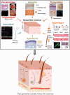
Figure 1
Figure 1
Ongoing studies towards building a complex human skin construct model. Top: Schematic of current skin constructs. Epidermis (Epi) is a stratified epithelium containing differentiated keratinocytes, which lies over the dermis made from fibroblasts mixed in a collagen matrix. Vascularization: (A) H&E and (B) immunofluorescent staining of histological sections of vascularized skin constructs generated using iPSC-derived ECs. The sections were immunolabeled with K14 (red), and CD31 (green) to evaluate epidermal integrity and endothelial coating in the microchannels. (C) The effect of vasculature pattern on the host neovascularization. Picture of newly formed host vasculature following the micropatterned human iEC-containing microchannels in vascularized skin constructs. Scale bars: 250 µm (with permission from Abaci et al.). Immunity: Immunohistochemical analysis of reconstructed epidermis containing Langerhans cells. Histochemical analysis of reconstructed epidermis containing Langerhans cells was performed using anti-Langerin staining (peroxidase/AEC reaction) with hematoxilin counterstaining was performed on cryosection to localize Langerhans cells inner the epidermal layers (the brown cells are the Langerin-positive cells) and Langerin immunostaining (B) on epidermal sheets visualized the dense network of the dendritic Langerhans cells. Scale bar: 50 µm (with permission from Facy et al.). Pigmentation: Skin constructs produced from iPSC-derived fibroblasts, keratinocytes, and melanocytes have normal anatomy and are functional. (A) Hematoxylin and eosin staining, (Continued) (B) Forskolin-treated iPSC-derived skin constructs after 14 days at the air–liquid interface. (C) Fontana-Masson and (D) Immunofluorescence of gp-100 (green) and nuclei (blue) staining in skin construct. Scale bars: 100 µm. (with permission from Gledhill et al.). Innervation: Detection of laminin and myelin sheaths in the reconstructed connective tissues enriched with Schwann cells. (A) Double immunofluorescent staining of neurons (160 kDa neurofilament, green) and Schwann cells (GFAP, red) on day 14. Schwann cells are colocalized with neurites (arrowheads). (B) Double immunofluorescent staining of neurons (160 kDa neurofilament, green) and laminin (red). The 160-kDa neurofilament staining colocalized with the laminin staining (arrowheads). Scale bars: 50 µm. (with permission from Blais et al.) Appendages: Transplantation of the bioengineered IOS. (A) Schematic representation of the methods used for the generation and transplantation of iPS cell-derived hair follicles. Cystic tissue with hair follicles was isolated and divided into small pieces containing 10 to 20 hair follicles. The small pieces were transplanted into the back skin of nude mice using a follicular unit transplantation (FUT) method developed in humans; (i) In vivo-organized integumentary organ system derived from male iPS cells (XY) (ii) Intra-cutaneous transplantation (iii) Orthotopic hair function. (B) Macromorphological observations of two independent engraftments into the dorsoventral skin of nude mice showing the eruption and growth of iPS cell-derived hair follicles. Scale bars: 1 mm. (C) Immunohistochemical analyses of stem/progenitor cells in the follicles of natural pelage and enhanced green fluorescent protein (EGFP)-labeled iPS cell-derived bioengineered hair follicles (with permission from Takagi et al.). (D) (i) Conventional culture systems enable the growth of cells in a 2D format. (ii) Growth of dermal papilla cells in hanging drop cultures results in the formation of dermal spheres. (iii) Alkaline phosphatase (AP) activity is not detected in passage four cultured dermal papilla cells. (iv) In dermal spheres, AP activity is strongly observed (with permission from Higgins et al.). Hypodermis: Full-thickness skin constructs using silk and collagen biomaterials. H&E of constructs cultured in 1:1 skin:adipose media, which had more physiologically relevant features (with permission from Bellas et al.). Bottom: Schematic of next-generation complex human skin constructs hosting all the different skin cell types as normal skin in vivo

References

    1. Fuchs E, Tumbar T, Guasch G. Socializing with the neighbors: stem cells and their niche. Cell 2004; 116: 769–78. - PubMed
    1. Millar SE. Molecular mechanisms regulating hair follicle development. J Invest Dermatol 2002; 118: 216–25. - PubMed
    1. Botchkarev VA, Paus R. Molecular biology of hair morphogenesis: development and cycling. J Exp Zool B Mol Dev Evol 2003; 298: 164–80. - PubMed
    1. Natarajan VT, Ganju P, Ramkumar A, Grover R, Gokhale RS. Multifaceted pathways protect human skin from UV radiation. Nat Chem Biol 2014; 10: 542–51. - PubMed
    1. Wang JX, Fukunaga-Kalabis M, Herlyn M. Crosstalk in skin: melanocytes, keratinocytes, stem cells, and melanoma. J Cell Commun Signal 2016; 10: 191–6. - PMC - PubMed

Publication types

MeSH terms