. 2017 Jul;19(7):747-757.
doi: 10.1177/1098612X17706471.

The feline blood film

Affiliations

The feline blood film

John W Harvey. J Feline Med Surg. 2017 Jul.

Abstract

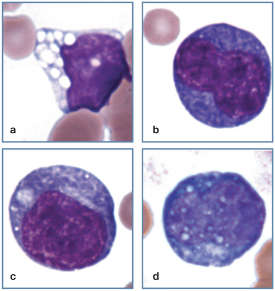
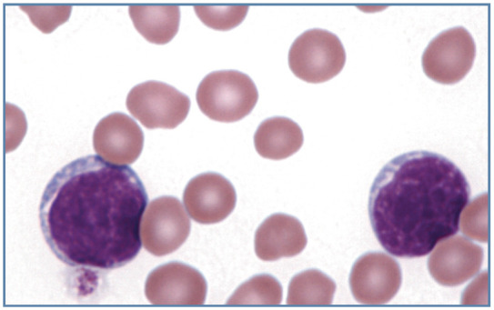
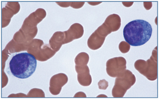
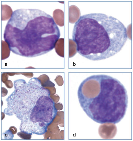
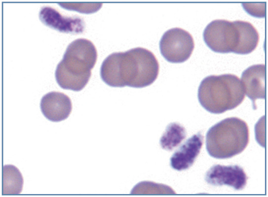
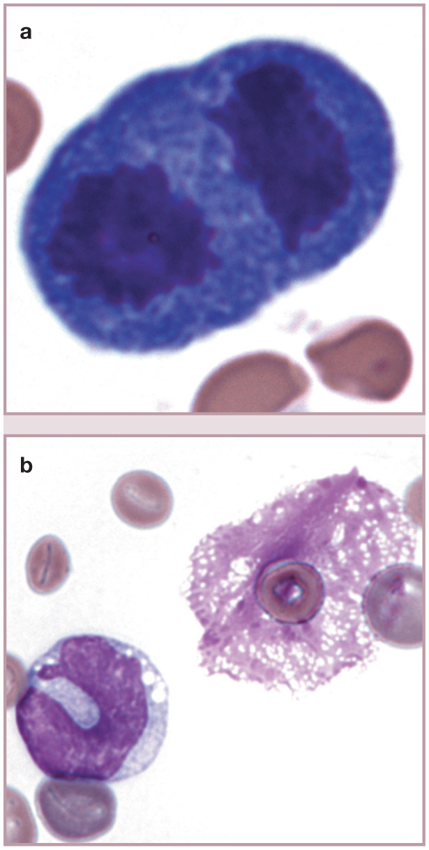
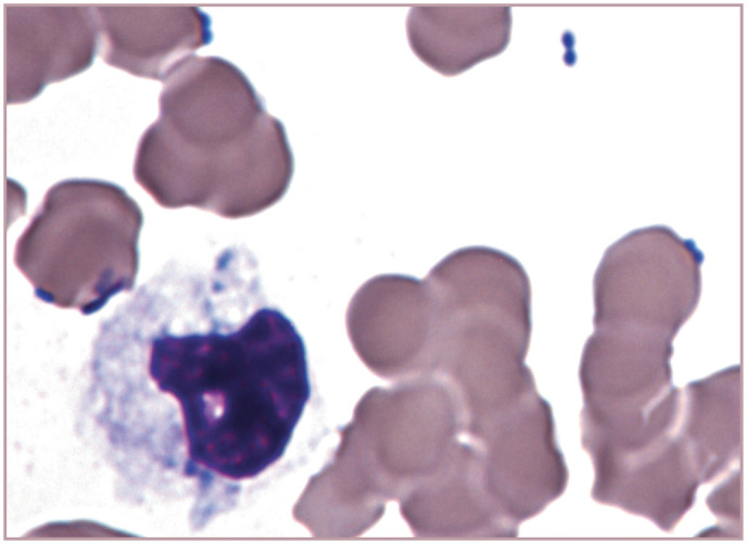
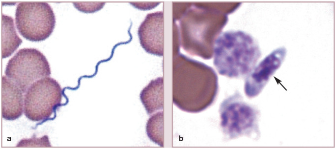
Practical relevance: Many veterinary practices have invested in quality automated hematology instruments for use in-house. However, regardless of their specific choice of analyzer, there are important hematology findings that can only be determined by microscopic examination of stained blood films. For this reason, and also for the purpose of quality control for the analyzer, a quick blood film review should be performed alongside every automated complete blood count. Even those practices that submit their blood samples to outside diagnostic laboratories for evaluation, still require the capability to examine stained blood films in emergency situations. Series outline: This is the second of a two-part article series that aims to familiarize the practitioner with normal findings on feline blood films, with a particular focus on unique features in the cat, as well as to assist with interpretation of common abnormalities. Part 2 focuses on the morphology of feline leukocytes and platelets in health and disease. Evidence base: The information and guidance offered is based on the published literature and the author's own extensive clinical pathology research.


Conflict of interest statement

The author declared no potential conflicts of interest with respect to the research, authorship, and/or publication of this article.

Figures

Figure 1
Figure 1
Morphology of feline neutrophils. (a) Normal neutrophil. (b) Band neutrophil. (c) Neutrophil from a female domestic medium hair cat. A Barr body (sex chromatin lobe or drumstick) is present (black arrow). Three nuclear vesicular appendages (blebs) are also present (one is marked by a red arrow); this cat was febrile and had suspected infection. Blood film courtesy of Matthew Williams. (d) Purple granules in the cytoplasm of an apparently healthy Siamese cat. Wright-Giemsa stain
Figure 2
Figure 2
Morphology of feline neutrophilic cells. (a) Large granules in the cytoplasm of a Persian cat with Chédiak- Higashi syndrome. (b) Neutrophil with Döhle bodies as the only evidence of cytoplasmic toxicity. (c) Large toxic neutrophil with foamy basophilic cytoplasm containing multiple darker blue Döhle bodies. Reproduced from Harvey (2012), with permission. (d) Band neutrophil with foamy basophilic cytoplasm containing Döhle bodies. Wright-Giemsa stain
Figure 3
Figure 3
Toxic left shift in blood from a cat with pyothorax. A band neutrophil (black arrow), early neutrophilic metamyelocyte (red arrow) and two neutrophils exhibit markedly basophilic cytoplasm. Wright-Giemsa stain
Figure 4
Figure 4
Toxic left shift in a cat with a leukemoid reaction (leukocyte count 101 × 106/l) resulting from a large draining abscess containing maggots. A toxic neutrophil (bottom left), toxic metamyelocyte (left), toxic myelocyte (top right), band neutrophil (right center) and neutrophil (bottom right) are present. Wright-Giemsa stain
Figure 5
Figure 5
Band neutrophil (left), bilobed neutrophil (center) and eosinophilic myelocyte (right) in the blood of a cat with Pelger-Huët anomaly. Wright-Giemsa stain. Reproduced from Harvey (2012), with permission
Figure 6
Figure 6
Abnormal neutrophil morphology. (a) Hypersegmented neutrophil in blood from a cat with myelodysplastic syndrome. Reproduced from Harvey (2012), with permission. (b) Toxic donut-shaped neutrophil and toxic giant band neutrophil in blood from a cat with pyothorax. Wright-Giemsa stain
Figure 7
Figure 7
Acute myeloid leukemia in a cat. Most round cells (myeloblasts and myelocytes) were peroxidase positive. A band neutrophil (bottom center) is also present. Wright-Giemsa stain
Figure 8
Figure 8
Blood from a cat with erythroleukemia (AML-M6). Large pale cells are predominantly myeloblasts. A basophilic rubriblast (black arrow) and a rubricyte with nuclear lobulation (red arrow) are also present. Wright-Giemsa stain
Figure 9
Figure 9
Feline neutrophils with phagocytized infectious organisms. (a) Bacterial rod in a neutrophil from a cat with a bacteremia. (b) Mycobacterium organisms in a neutrophil; these do not stain and appear as clear linear structures. (c) Hepatozoon felis gamont in a neutrophil. Reproduced from Lloret et al (2015), with permission. Image courtesy of G Baneth. (d) Leishmania amastigote in a feline neutrophil. Reproduced from Harvey (2012), with permission. Image courtesy of M Santos
Figure 10
Figure 10
Feline eosinophils containing rod-shaped granules. (a) Bilobed mature eosinophil. (b) Band eosinophil. Wright-Giemsa stain
Figure 11
Figure 11
Feline basophils. (a) Basophil containing many light lavender granules in the cytoplasm. (b) Basophil with a mixture of light-lavender and purple granules. The pale granules overlying the nucleus give it a moth-eaten appearance. Wright-Giemsa stain
Figure 12
Figure 12
Normal lymphocyte morphology. (a) Small lymphocyte. (b) Medium-sized lymphocyte. (c) Large lymphocyte. (d) Granular lymphocyte. Wright-Giemsa stain
Figure 13
Figure 13
Two neoplastic large granular lymphocytes in blood from a cat with metastatic large granular lymphoma. Granules are larger than those seen in non-neoplastic granular lymphocytes. Wright-Giemsa stain
Figure 14
Figure 14
Abnormal lymphocyte morphology. (a) Vacuolated lymphocyte from a Korat cat with inherited GM2 gangliosidosis. (b,c) Reactive lymphocytes with increased cytoplasmic basophilia. (d) Reactive lymphocyte with cytoplasmic Russell bodies containing immunoglobulin. Wright-Giemsa stain
Figure 15
Figure 15
Blood film from a cat with acute lymphoblastic leukemia, showing two large lymphoblasts. Wright-Giemsa stain
Figure 16
Figure 16
Neoplastic plasma cells in the blood of a cat with a metastatic plasma cell neoplasm involving bone marrow. There is increased basophilia between cells and prominent rouleaux formation because a monoclonal hyperglobulinemia was present. Wright-Giemsa stain
Figure 17
Figure 17
Mononuclear phagocyte morphology. (a,b) Normal monocyte morphology. (c) Macrophage at the feathered end of a blood film from a cat with Mycoplasma haemofelis infection. The magnification of this image is less than others in this group. (d) Monocyte with a phagocytized erythrocyte in blood from a cat with M haemofelis infection. Wright-Giemsa stain
Figure 18
Figure 18
Disrupted intermediate Cytauxzoon felis schizont at the feathered end of a blood film. The large round magenta nucleus containing a blue nucleolus is from the host macrophage. Wright–Giemsa stain
Figure 19
Figure 19
Mast cell (left) and basophil (right) in blood from a cat with splenic mast cell neoplasia. The round nucleus of the mast cell is nearly obscured by the large numbers of cytoplasmic granules present. Wright-Giemsa stain
Figure 20
Figure 20
Morphology of platelets. (a) Aggregate demonstrating the large size and variable size of feline platelets. The platelet at the bottom is larger than seen in most cats. (b) Macroplatelet in blood from a cat with erythroleukemia. Wright-Giemsa stain
Figure 21
Figure 21
Aggregate of largely degranulated platelets. Wright-Giemsa stain
Figure 22
Figure 22
Large elongated platelets from a Ragdoll cat that was homozygous for the F385Y change in the gene encoding for β1-tubulin. Wright-Giemsa stain. Courtesy of EA Spangler
Figure 23
Figure 23
Mitotic figure and lysed cell. (a) A cell in telophase of division in blood from a cat with M haemofelis infection and regenerative anemia. The cell was most likely a lymphocyte based on the cytoplasmic basophilia, but an early nucleated erythroid precursor is also possible. (b) Monocyte (left) and free nucleus from a lysed cell (right). Nuclei with this net-like appearance have been called basket cells, even though they are not intact cells. Wright-Giemsa stain
Figure 24
Figure 24
Bacteria free in the plasma of a cat with marked leukopenia and septic shock. A toxic karyolytic neutrophil is present. Harleco Fucillo stain. Photograph from a stained blood film from a 2014 ASVCP slide review submitted by JE Stickle
Figure 25
Figure 25
Infectious organisms free in plasma. (a) Relapsing fever spirochete (Borrelia persica) in blood from a cat. Courtesy of G Baneth. (b) A Sarcocystis species tachyzoite in the plasma of a cat (arrow). Two platelets and multiple erythrocytes are present. Wright-Giemsa stain. Photograph from a stained blood film from a 2015 ASVCP slide review submitted by N Zitzer, MJ Radin, M Wellman and A Marsh
Figure 26
Figure 26
Dirofilaria immitus microfilaria in blood from a cat with heartworm disease. Wright-Giemsa stain. Reproduced from Harvey (2012), with permission
None
None
None
None

References

    1. Harvey JW. Veterinary hematology. A diagnostic guide and color atlas. St Louis, MO: Elsevier Saunders, 2012.
    1. Bessis M. Living blood cells and their ultrastructure. New York: Springer-Verlag, 1973, pp 337–338.
    1. Bunting RW, Selig MK. Localization of DNA in ultrascopic nuclear appendages of polymorphonuclear white blood cells from patients with low serum B(12). J Histochem Cytochem 2002; 50: 1381–1388. - PubMed
    1. Hirsch VM, Cunningham TA. Hereditary anomaly of neutrophil granulation in Birman cats. Am J Vet Res 1984; 45: 2170–2174. - PubMed
    1. Kramer JW, Davis WC, Prieur DJ. The Chédiak-Higashi syndrome of cats. Lab Invest 1977; 36: 554–562. - PubMed

LinkOut - more resources