

Angiostrongylus vasorum infection in dogs from a cardiopulmonary dirofilariosis endemic area of Northwestern Italy: a case study and a retrospective data analysis
- PMID: 28592295
- PMCID: PMC5463301
- DOI: 10.1186/s12917-017-1083-7
Angiostrongylus vasorum infection in dogs from a cardiopulmonary dirofilariosis endemic area of Northwestern Italy: a case study and a retrospective data analysis
Abstract
Background: In Italy, Angiostrongylus vasorum, an emergent parasite, is being diagnosed in dogs from areas considered free of infection so far. As clinical signs are multiple and common to other diseases, its diagnosis can be challenging. In particular, in areas where angiostrongylosis and dirofilariosis overlap, a misleading diagnosis of cardiopulmonary dirofilariosis might occur even on the basis of possible misleading outcomes from diagnostic kits.
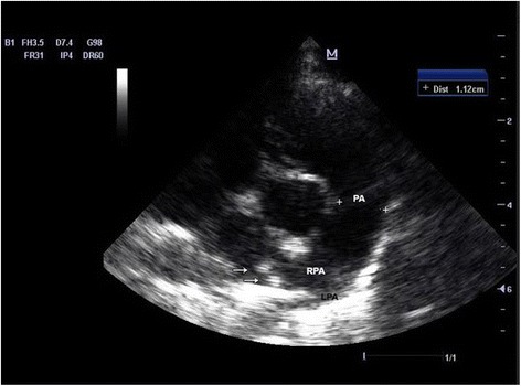
Case presentation: Two Cavalier King Charles spaniel dogs from an Italian breeding in the Northwest were referred to a private veterinary hospital with respiratory signs. A cardiopulmonary dirofilariosis was diagnosed and the dogs treated with ivermectin, but one of them died. At necropsy, pulmonary oedema, enlargement of tracheo-bronchial lymphnodes and of cardiac right side were detected. Within the right ventricle lumen, adults of A. vasorum were found. All dogs from the same kennel were subjected to faecal examination by FLOTAC and Baermann's techniques to detect A. vasorum first stage larvae; blood analysis by Knott's for Dirofilaria immitis microfilariae, and antigenic tests for both A. vasorum (Angio Detect™) and D.immitis (DiroCHEK® Heartworm, Witness®Dirofilaria). The surviving dog with respiratory signs resulted positive for A. vasorum both at serum antigens and larval detection. Its Witness® test was low positive similarly to other four dogs from the same kennel, but false positive results due to cross reactions with A. vasorum were also considered. No dogs were found infected by A. vasorum. Eventually, the investigation was deepened by browsing the pathological database of Veterinary Pathology Laboratories at Veterinary School of Milan University through 1998-2016, where 11 cases of angiostrongylosis were described. Two out of 11 dogs had a mixed infection with Crenosoma vulpis.
Conclusion: The study demonstrates the need for accurate surveys to acquire proper epidemiological data on A. vasorum infection in Northwestern Italy and for appropriate diagnostic methods. Veterinary clinicians should be warned about the occurrence of this canine parasite and the connected risk of a misleading diagnosis, particularly in areas endemic for cardiopulmonary dirofilariosis.
Keywords: Angio detect™; Angiostrongylus vasorum; Crenosoma vulpis; DiroCHEK® heartworm; Dirofilaria immitis; Dogs; FLOTAC; Witness® Dirofilaria.
Figures


References
-
- Guilhon J, Cens B. Angiostrongylus vasorum (Baillet, 1866) étude biologique et morphologique. Ann Parasitol Hum Comp. 1973;48:567–596. - PubMed
-
- Serres E. Entozoaires trouvés dans l’oreille droite, le ventricule correspondant et l'artère pulmonaire d'un chien. J Vétérinaires Midi. 1854;7:70.
Publication types
MeSH terms
LinkOut - more resources
Full Text Sources
Other Literature Sources
Medical
Research Materials
