

Machine learning techniques for diabetic macular edema (DME) classification on SD-OCT images
- PMID: 28592309
- PMCID: PMC5463338
- DOI: 10.1186/s12938-017-0352-9
Machine learning techniques for diabetic macular edema (DME) classification on SD-OCT images
Abstract
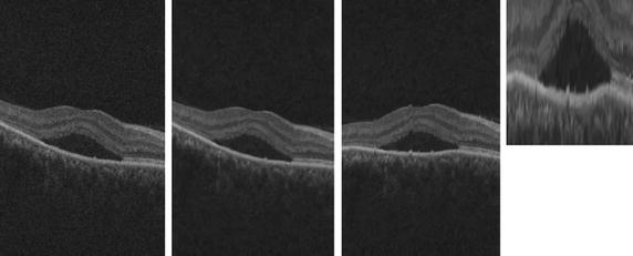
Background: Spectral domain optical coherence tomography (OCT) (SD-OCT) is most widely imaging equipment used in ophthalmology to detect diabetic macular edema (DME). Indeed, it offers an accurate visualization of the morphology of the retina as well as the retina layers.
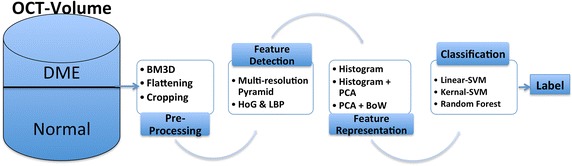
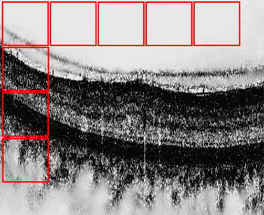
Methods: The dataset used in this study has been acquired by the Singapore Eye Research Institute (SERI), using CIRRUS TM (Carl Zeiss Meditec, Inc., Dublin, CA, USA) SD-OCT device. The dataset consists of 32 OCT volumes (16 DME and 16 normal cases). Each volume contains 128 B-scans with resolution of 1024 px × 512 px, resulting in more than 3800 images being processed. All SD-OCT volumes are read and assessed by trained graders and identified as normal or DME cases based on evaluation of retinal thickening, hard exudates, intraretinal cystoid space formation, and subretinal fluid. Within the DME sub-set, a large number of lesions has been selected to create a rather complete and diverse DME dataset. This paper presents an automatic classification framework for SD-OCT volumes in order to identify DME versus normal volumes. In this regard, a generic pipeline including pre-processing, feature detection, feature representation, and classification was investigated. More precisely, extraction of histogram of oriented gradients and local binary pattern (LBP) features within a multiresolution approach is used as well as principal component analysis (PCA) and bag of words (BoW) representations.
Results and conclusion: Besides comparing individual and combined features, different representation approaches and different classifiers are evaluated. The best results are obtained for LBP[Formula: see text] vectors while represented and classified using PCA and a linear-support vector machine (SVM), leading to a sensitivity(SE) and specificity (SP) of 87.5 and 87.5%, respectively.
Keywords: BoW; Classification; DME detection; HoG; LBP; SD-OCT.
Figures
Similar articles
-
Classification of SD-OCT volumes with multi pyramids, LBP and HOG descriptors: application to DME detections.Annu Int Conf IEEE Eng Med Biol Soc. 2016 Aug;2016:1344-1347. doi: 10.1109/EMBC.2016.7590956. Annu Int Conf IEEE Eng Med Biol Soc. 2016. PMID: 28268574
-
Fully Automated Detection and Quantification of Macular Fluid in OCT Using Deep Learning.Ophthalmology. 2018 Apr;125(4):549-558. doi: 10.1016/j.ophtha.2017.10.031. Epub 2017 Dec 8. Ophthalmology. 2018. PMID: 29224926
-
Evaluation of an Artificial Intelligence-Based Detector of Sub- and Intraretinal Fluid on a Large Set of Optical Coherence Tomography Volumes in Age-Related Macular Degeneration and Diabetic Macular Edema.Ophthalmologica. 2022;245(6):516-527. doi: 10.1159/000527345. Epub 2022 Oct 10. Ophthalmologica. 2022. PMID: 36215958
-
Pathological Neurovascular Unit Mapping onto Multimodal Imaging in Diabetic Macular Edema.Medicina (Kaunas). 2023 May 7;59(5):896. doi: 10.3390/medicina59050896. Medicina (Kaunas). 2023. PMID: 37241128 Free PMC article. Review.
-
Role of Inflammation in Classification of Diabetic Macular Edema by Optical Coherence Tomography.J Diabetes Res. 2019 Dec 20;2019:8164250. doi: 10.1155/2019/8164250. eCollection 2019. J Diabetes Res. 2019. PMID: 31930145 Free PMC article. Review.
Cited by
-
Optical coherence tomography image based eye disease detection using deep convolutional neural network.Health Inf Sci Syst. 2022 Jun 21;10(1):13. doi: 10.1007/s13755-022-00182-y. eCollection 2022 Dec. Health Inf Sci Syst. 2022. PMID: 35756852 Free PMC article.
-
Automated OCT angiography image quality assessment using a deep learning algorithm.Graefes Arch Clin Exp Ophthalmol. 2019 Aug;257(8):1641-1648. doi: 10.1007/s00417-019-04338-7. Epub 2019 May 22. Graefes Arch Clin Exp Ophthalmol. 2019. PMID: 31119426
-
Detection and Classification of Diabetic Macular Edema with a Desktop-Based Code-Free Machine Learning Tool.Turk J Ophthalmol. 2023 Oct 19;53(5):301-306. doi: 10.4274/tjo.galenos.2023.92635. Turk J Ophthalmol. 2023. PMID: 37868586 Free PMC article.
-
Artificial intelligence in retinal disease: clinical application, challenges, and future directions.Graefes Arch Clin Exp Ophthalmol. 2023 Nov;261(11):3283-3297. doi: 10.1007/s00417-023-06052-x. Epub 2023 May 9. Graefes Arch Clin Exp Ophthalmol. 2023. PMID: 37160501 Free PMC article. Review.
-
Autonomous Screening for Diabetic Macular Edema Using Deep Learning Processing of Retinal Images.Ophthalmol Sci. 2025 Jan 31;5(4):100722. doi: 10.1016/j.xops.2025.100722. eCollection 2025 Jul-Aug. Ophthalmol Sci. 2025. PMID: 40225408 Free PMC article.
References
MeSH terms
LinkOut - more resources
Full Text Sources
Other Literature Sources
Medical
Miscellaneous
