Comparative Study
. 2017 Aug 4;292(31):13056-13067.
doi: 10.1074/jbc.M117.787192. Epub 2017 Jun 7.

Exploiting fine-scale genetic and physiological variation of closely related microbes to reveal unknown enzyme functions

Affiliations
Comparative Study

Exploiting fine-scale genetic and physiological variation of closely related microbes to reveal unknown enzyme functions

Ahmet H Badur et al. J Biol Chem. .

Abstract

Polysaccharide degradation by marine microbes represents one of the largest and most rapid heterotrophic transformations of organic matter in the environment. Microbes employ systems of complementary carbohydrate-specific enzymes to deconstruct algal or plant polysaccharides (glycans) into monosaccharides. Because of the high diversity of glycan substrates, the functions of these enzymes are often difficult to establish. One solution to this problem may lie within naturally occurring microdiversity; varying numbers of enzymes, due to gene loss, duplication, or transfer, among closely related environmental microbes create metabolic differences akin to those generated by knock-out strains engineered in the laboratory used to establish the functions of unknown genes. Inspired by this natural fine-scale microbial diversity, we show here that it can be used to develop hypotheses guiding biochemical experiments for establishing the role of these enzymes in nature. In this work, we investigated alginate degradation among closely related strains of the marine bacterium Vibrio splendidus One strain, V. splendidus 13B01, exhibited high extracellular alginate lyase activity compared with other V. splendidus strains. To identify the enzymes responsible for this high extracellular activity, we compared V. splendidus 13B01 with the previously characterized V. splendidus 12B01, which has low extracellular activity and lacks two alginate lyase genes present in V. splendidus 13B01. Using a combination of genomics, proteomics, biochemical, and functional screening, we identified a polysaccharide lyase family 7 enzyme that is unique to V. splendidus 13B01, secreted, and responsible for the rapid digestion of extracellular alginate. These results demonstrate the value of querying the enzymatic repertoires of closely related microbes to rapidly pinpoint key proteins with beneficial functions.

Keywords: Vibrio splendidus; algae; alginate lyase; bacteria; enzyme kinetics; mass spectrometry (MS); nuclear magnetic resonance (NMR).


Conflict of interest statement

The authors declare that they have no conflicts of interest with the contents of this article

Figures

Figure 1.
Figure 1.
Comparison of secreted alginate lyase activity among closely related V. splendidus strains. A, phylogenetic comparison of closely related V. splendidus strains. The species tree was calculated as described previously with concatenated ribosomal proteins (21). Scale bar, substitutions per site. Notably, all of these strains can catabolize alginate (21). B, alginate lyase secretion assay on agar plates with marine broth medium supplemented with 0.25% alginate. The Vibrio strains were grown for 36 h, and the resulting colonies were imaged. Cetylpyridinium chloride revealed the dark halos indicative of alginate (opaque background) that was digested by secreted alginate lyase diffusing beyond the colony boundaries. The bars (blue; mean) and error bars (S.D.) were calculated from the four technical replicates shown in the left panel.
Figure 2.
Figure 2.
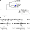
Alginate lyases in V. splendidus 12B01 and 13B01. A, rooted maximum likelihood tree of PL7 lyases in V. splendidus 12B01 and 13B01. The PL7 enzyme from Alteromonas sp. 272 was used as an outgroup. The numbers at nodes represent bootstrap values derived from 100 tree calculations. B, domain structure of alginate lyases in V. splendidus 13B01. The indicated amino acids are the hypothesized catalytic residues. CBM32 is the carbohydrate binding module family 32 domain. The small flags indicate signal peptides, where the white and black boxes are used specify whether these peptides are predicted to be cleaved by signal peptidase I or II, respectively. The signal peptides of secreted lyases are expected to be cleaved by signal peptidase I, and the signal peptides of membrane-associated lyases are expected to be cleaved by signal peptidase II (25, 49). The GenBankTM accession numbers are as follows: PL7A (OCH64533.1), PL7B (OCH64532.1), PL7D (OCH64519.1), PL7E (OCH62672.1), PL6F (OCH64519.1), and PL7G (OCH63958.1).
Figure 3.
Figure 3.
Expression of alginate lyases in V. splendidus 12B01 and 13B01 during growth on glucose and alginate as determined using quantitative PCR. The housekeeping gene rpoA was used as an internal control. The data for V. splendidus 12B01 were published previously (22) and are included for comparative purposes. Bars, mean of three technical replicates; error bars, S.D.
Figure 4.
Figure 4.
Comparison of alginate lyase expression and localization in V. splendidus 12B01 and 13B01 as determined by proteomics. Bars, mean of three technical replicates; error bars, S.D. The data are presented as NSAFs.
Figure 5.
Figure 5.
1H NMR (400-MHz) spectra of alginate and alginate-derived substrates following degradation with PL7A, PL7B, PL7D, PL7E, PL6F, and PL7G. G and M, signals from internal G and M residues, respectively; G-beta and M-beta, signals from reducing G and M residues, respectively; Δ, signal from 4-deoxy-l-erythro-hex-4-enepyranosyluronate non-reducing end residue. Non-underlined residues are the neighboring residues to those generating each signal. Protons (H) are numbered to indicate which particular proton causes the signal. A, alginate; B, poly-G–enriched alginate; C, poly-M–enriched alginate.
Figure 6.
Figure 6.
Negative-ion electrospray ionization mass spectra of alginate lyase-degraded alginate. DP and the respective integers (1–6) refer to the degree of polymerization. m/z values are listed below the DP values. Although the ESI-MS analysis of PL7A (A) only detected monomeric and dimeric degradation products, NMR shows that the actual product size has an average DP of 17 (data not shown). These longer-chain substrates are not detected by ESI-MS due to low signal intensity. The other lyases (B–F) are clearly endolytic, because ESI-MS analysis shows a ladder-type range of degradation products that is common for endo-acting enzymes degrading gel-forming, algal polysaccharides (9).
Figure 7.
Figure 7.
Comparison of secreted alginate lyase activities in wild-type V. splendidus 12B01 and 13B01 and a V. splendidus 13B01 Δpl7G mutant. The top three rows show the colony images for the respective strains grown on a marine broth medium agar plate supplemented with 0.25% alginate. The bottom three rows show secreted alginate lyase activity of the same colonies, after they were manually removed, as determined by staining with cetylpyridinium chloride.

References

    1. Falkowski P. G., Barber R. T., and Smetacek V. V. (1998) Biogeochemical controls and feedbacks on ocean primary production. Science 281, 200–207 - PubMed
    1. Alderkamp A. C., van Rijssel M., and Bolhuis H. (2007) Characterization of marine bacteria and the activity of their enzyme systems involved in degradation of the algal storage glucan laminarin. FEMS Microbiol. Ecol. 59, 108–117 - PubMed
    1. Torode T. A., Marcus S. E., Jam M., Tonon T., Blackburn R. S., Hervé C., and Knox J. P. (2015) Monoclonal antibodies directed to fucoidan preparations from brown algae. PLoS One 10, e0118366. - PMC - PubMed
    1. Myklestad S. M. (1995) Release of extracellylar products by phtyoplankton with special emphasis on polysaccharides. Sci. Total Environ. 165, 155–164
    1. Martin J. H., Knauer G. A., Karl D. M., and Broenkow W. W. (1987) VERTEX: carbon cycling in the northeast Pacific. Deep Sea Res. A 34, 267–285

Publication types

MeSH terms

LinkOut - more resources