Review
. 2017 May;19(2):131-142.
doi: 10.5853/jos.2017.00283. Epub 2017 May 31.

Causes and Solutions of Endovascular Treatment Failure

Affiliations
Review

Causes and Solutions of Endovascular Treatment Failure

Byung Moon Kim. J Stroke. 2017 May.

Abstract

In a meta-analysis of individual patient data from 5 randomized controlled trials, endovascular treatment (EVT) mainly using a stent retriever achieved successful recanalization in 71.1% of patients suffering from acute stroke due to anterior circulation large artery occlusion (LAO). However, EVT still failed in 28.9% of LAO cases in those 5 successful trials. Stent retriever failure may occur due to anatomical challenges (e.g., a tortuous arterial tree from the aortic arch to a target occlusion site), a large quantity of clots, tandem occlusion, clot characteristics (fresh versus organized clots), different pathomechanisms (embolic versus non-embolic occlusion), etc. Given that recanalization success is the most important factor in the neurological outcome of acute stroke patients, it is important to seek solutions for such difficult cases. In this review, the basic technique of EVT is briefly summarized and then various difficult cases with diverse conditions are discussed along with suggested solutions.

Keywords: Acute stroke; Endovascular thrombectomy; Stent retriever.


Conflict of interest statement

The author has no conflicts of interest.

Figures

Figure 1.
Figure 1.
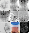
A 60-year-old man presented with left side weakness (initial NIHSS score of 15). (A) A right carotid angiogram revealed occlusion of right internal carotid artery at the proximal cervical segment. (B) The delayed phase of the angiogram disclosed tandem occlusion of the right internal carotid artery at the supraclinoid segment. (C) After a balloon-guiding catheter was placed in the common carotid artery, the internal carotid artery was navigated using a microcatheter (arrowheads). (D) After an angioplasty balloon catheter was advanced over the 300-cm length exchangeable microwire, a balloon angioplasty was performed. (E) A closed-cell carotid stent was placed while the distal tip of the balloon of the guiding catheter was inflated (arrow). (F) A balloon-guiding catheter was advanced to the end of the carotid stent. Thereafter, the angiogram shows a tandem occlusion of the supraclinoid internal carotid artery. (G) A microcatheter was navigated into the middle cerebral artery branch during balloon inflation (arrow). (H) A Solitaire stent retriever was deployed spanning the entire clot length. A curved arrow indicates a distal marker of the deployed Solitaire. Arrow indicates inflated balloon guide catheter. (I) A large amount of clots was removed with the Solitaire through the balloon-guiding catheter suction. (J) Control angiogram revealed complete (modified Thrombolysis in Cerebral Ischemia [mTICI] 3) recanalization. (K) Diffusion-weighted magnetic resonance imaging on the following day showed small areas of acute infarctions in the right middle cerebral artery territory. NIHSS, the National Institutes of Health Stroke Scale.
Figure 2.
Figure 2.
A 74-year-old woman presented with mental changes and right side weakness (initial NIHSS score of 18). (A) A right carotid angiogram showed left internal carotid artery terminal occlusion. (B) A spot image during the navigation of an angiocatheter disclosed the left common carotid artery at the proximal occlusion (curved arrow). (C) After suction of a large quantity of clots through the 8F shuttle sheath (long arrow), an 8F balloonguiding catheter was advanced up to the carotid bulb (short arrow). Another suction was performed through the balloon-guiding catheter after inflation of the tip of the balloon. Note the large quantity of clots (filling defects, arrowheads). (D) After removal of the balloon-guiding catheter because it was occluded with packed clots, the shuttle sheath was also found to be occluded with clots (arrowheads). (E) After forced suction through shuttle sheath, an angiogram revealed occlusion of an internal carotid artery cavernous segment. The balloon-guiding catheter (short arrow) was reintroduced within a shuttle sheath (long arrow) and advanced up to a high cervical segment. (F) The left middle cerebral artery main branch was navigated using a microcatheter (white arrow). (G) After a Solitaire stent retriever was passed once, the control angiogram showed complete recanalization. (H) A large number of clots in the bottle. Most of the clots were removed through suction of the shuttle sheath and the balloonguiding catheter. (I) Diffusion-weighted magnetic resonance imaging on the following day disclosed scattered acute infarctions in the left middle cerebral artery. NIHSS, the National Institutes of Health Stroke Scale.
Figure 3.
Figure 3.
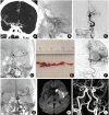
A 47-year-old man presented with aphasia and right side weakness (initial NIHSS score of 21). (A) An initial computed tomography angiogram showed occlusion of the left middle cerebral artery orifice. (B) A spot image during the navigation of a balloon-guiding catheter showed an occlusion of the high cervical portion of the internal carotid artery. (C) After a single suction thrombectomy through a balloon-guiding catheter, an angiogram showed a dissection (arrows) of the high cervical segment of the internal carotid artery with a tandem occlusion in the middle of the cerebral artery orifice. (D) For bypass of the tortuous and dissected segment of the internal carotid artery (dotted circle), a 6F intermediate catheter (arrowhead) was advanced after deployment of a Solitaire (4×20 mm), which served as an anchor during the advancement of the intermediate catheter. A curved arrow indicates the distal markers of the Solitaire stent retriever. (E) A long clot retrieved with the Solitaire. (F) A control angiogram showed complete recanalization of the left middle cerebral artery. (G) Another Solitaire stent (6×30 mm) was placed and detached, spanning the dissected segment (curved arrows) of the internal carotid artery. (H) Diffusion-weighted magnetic resonance imaging on the following day showed acute infarction in the left basal ganglia, insula, and opercular portion. (I) A contrast-enhanced magnetic resonance angiogram showed a patent left internal carotid artery through the stented segment (curved arrows). NIHSS, the National Institutes of Health Stroke Scale.
Figure 4.
Figure 4.
A 65-year-old woman presented with left side weakness (initial NIHSS score of 17). (A) A computed tomography angiogram showed an occlusion of the right internal carotid artery terminal portion (long arrow). (B) On the same computed tomography angiogram, take-off of the right posterior cerebral artery from the basilar artery was not visualized (short arrow), suggestive of a fetal-type posterior cerebral artery arising from internal carotid artery. (C) A right carotid angiogram showed an occlusion of the right internal carotid artery petro-cavernous segment. Note the tortuosity of the cervical segment (dotted circle) of the internal carotid artery. (D) After deployment of a Solitaire, a 6F intermediate catheter (arrowhead) was advanced facing the clots. A curved arrow indicates the distal markers of the deployed Solitaire. (E) A 3-minute delay angiogram showed long clots from the cavernous segment of the internal carotid artery to the middle cerebral artery. (F) A large quantity of clots was removed with the Solitaire and through the intermediate catheter. (G, H) An anteroposterior and lateral control angiogram showed complete recanalization. Note the fetal-type posterior cerebral artery (short arrow) originating from the internal carotid artery. (I) Diffusion-weight magnetic resonance imaging on the following day showed a large area of cortical infarction along the middle cerebral and posterior cerebral artery areas. NIHSS, the National Institutes of Health Stroke Scale.
Figure 5.
Figure 5.
A schematic comparing a branching-site (embolic, left) occlusion and a truncal-type (nonembolic, right) occlusion.
Figure 6.
Figure 6.
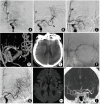
A 67-year-old man presented with aphasia and right side weakness (initial NIHSS score of 13). (A) A left carotid angiogram showed left middle cerebral artery M1 occlu - sion. (B) A control angiogram just after a Soli - taire deployment showed complete recanaliza - tion. An arrowhead indicates the distal markers of the Solitaire. (C) An angiogram after retrieval of the Solitaire showed re-occlusion. This phe - nomenon of complete recanalization after the Solitaire placement and re-occlusion after the retrieval was repeated 5 times. (D) A 3D vol - ume-rendering reconstruction image obtained during the Solitaire deployment showed 2 spots of stenosis (white arrows). Note the bifurcation (curved arrow) of the middle cerebral artery that was saved. (E) A flat-panel angiographic computed tomography showed minimal con - trast agent enhancement in the left middle ce - rebral artery territory. A glycoprotein IIb/IIIa in - hibitor was intra-arterially administered through the microcatheter, but the occlusion showed no response. (F) A Wingspan stent was placed after the balloon angioplasty. (G) The fi - nal control angiogram after permanent stenting showed complete recanalization. (H) Diffusionweighted magnetic resonance imaging on the following day showed several small high signal spots in the left middle cerebral artery area. (I) A 3-month follow-up computed tomography angiogram showed that the stented segment of the left middle cerebral artery was patent. NI - HSS, the National Institutes of Health Stroke Scale.
Figure 7.
Figure 7.
A 58-year-old woman presented with left side weakness (initial NIHSS score of 14). (A) A right carotid angiogram showed an occlusion of the right middle cerebral artery orifice. (B) After a single Solitaire pass, the M1 segment and inferior division (arrow) were recanalized, but the superior division was still occluded. (C) After 2 more Solitaire passes, the superior divi - sion was still occluded and showed a spastic appearance. A 4th Solitaire pass was performed after intra-arterial vasodilator (nimodipin, 0.5 mg) infusion (arrow) through the microcatheter while the Solitaire was deployed. (D) Two orga - nized clots from M1 and M2 (superior division). (E) The control angiogram after the 4th pass of the Solitaire showed complete recanalization of the middle cerebral artery. (F) Diffusion-weight - ed magnetic resonance imaging on the follow - ing day showed small areas of acute infarction in the right middle cerebral artery territory. NI - HSS, the National Institutes of Health Stroke Scale.
Figure 8.
Figure 8.
A 75-year-old woman presented with left side weakness (initial NIHSS score of 15). (A) A spot image during the navigation of a balloon-guiding catheter showed an occlusion of the right cervical internal carotid artery. (B) Suction thrombectomy was performed once through the balloon guiding catheter (arrowhead). (C) A control angiogram after suction thrombectomy showed right middle cerebral artery distal occlusion. (D) After a single Solitaire pass, partial recanalization was achieved. (E) In spite of repeated Solitaire passes, one clot (filling defect, arrow) was not removed. (F) A 5F intermediate catheter was advanced facing the clot (arrow) after anchoring the Solitaire (curved arrow). (G) The Solitaire retrieval yielded nothing, but the organized clot was captured by the intermediate catheter. (H) The final control angiogram showed complete recanalization. (I) Diffusion-weighted magnetic resonance imaging on the following day showed two high signal spots in the right middle cerebral artery territory. NIHSS, the National Institutes of Health Stroke Scale.
Figure 9.
Figure 9.
Schematic drawings show interactions of the stent with soft (red blood cell-rich) clots or organized (fibrin-rich) clots.
Figure 10.
Figure 10.
A 72-year-old woman presented with aphasia and right side weakness (initial NIHSS score of 19). (A) The lowest maximum intensity projection reconstruction image from a cerebral computed tomography angiogram showed a pedunculated thrombus in the left atrium. (B) A left carotid angiogram showed left middle cerebral artery M1 occlusion. (C) An angiogram with Solitaire in situ showed partial recanalization. (D) An angiogram after 5 passes of the Solitaire showed partial recanalization of the anterior temporal artery. (E) The clots were removed after 5 passes of the Solitaire. Note that the clots are resilient rather than sticky, suggestive of organized clots. (F) Suction thrombectomy was attempted using a Penumbra catheter (arrow). (G) An angiogram after 3 suction thrombectomy passes still showed occlusion in the middle cerebral artery beyond the orifice of the anterior temporal artery. (H) A flat-panel angiographic computed tomography showed minimal contrast agent enhancement in the left middle cerebral artery territory. Thereafter, a glycoprotein IIb/IIIa inhibitor was administered, but the occlusion showed no response. (I) A 30-minute delay angiogram showed a patent left middle cerebral artery while the Solitaire was deployed but not detached. (J) The same angiogram showed at least modified TICI 2b recanalization. Therefore, the Solitaire stent was detached. (K) Diffusion-weighted magnetic imaging on the following day showed scattered small infarctions in the left middle cerebral artery territory. (L) A magnetic resonance angiogram on the following day showed patent left middle cerebral artery branches (curved arrow). Note that the left middle cerebral artery trunk is not seen due to a metallic artifact of the stent. NIHSS, the National Institutes of Health Stroke Scale.

References

    1. Berkhemer OA, Fransen PS, Beumer D, van den Berg LA, Lingsma HF, Yoo AJ, et al. A randomized trial of intraarterial treatment for acute ischemic stroke. N Engl J Med. 2015;372:11–20. - PubMed
    1. Campbell BCV, Mitchell PJ, Kleinig TJ, Dewey HM, Churilov L, Yassi N, et al. Endovascular therapy for ischemic stroke with perfusion-imaging selection. N Engl J Med. 2015;372:1009–1018. - PubMed
    1. Goyal M, Demchuk AM, Menon BK, Eesa M, Rempel JL, Thornton J, et al. Randomized assessment of rapid endovascular treatment of ischemic stroke. N Engl J Med. 2015;372:1019–1030. - PubMed
    1. Jovin TG, Chamorro A, Cobo E, de Miguel MA, Molina CA, Rovira A, et al. Thrombectomy within 8 hours after symptom onset in ischemic stroke. N Engl J Med. 2015;372:2296–2306. - PubMed
    1. Saver JL, Goyal M, Bonafe A, Diener HC, Levy EI, Pereira VM, et al. Stent-retriever therombectomy after intravenous t-PA vs. t-PA alone in stroke. N Engl J Med. 2015;372:2285–2295. - PubMed