Review
. 2017 May;19(2):143-151.
doi: 10.5853/jos.2017.00626. Epub 2017 May 31.

Diagnostic and Therapeutic Strategies for Acute Intracranial Atherosclerosis-related Occlusions

Affiliations
Review

Diagnostic and Therapeutic Strategies for Acute Intracranial Atherosclerosis-related Occlusions

Jin Soo Lee et al. J Stroke. 2017 May.

Abstract

Intracranial atherosclerosis-related occlusion (ICAS-O) is frequently encountered at the time of endovascular revascularization treatment (ERT), especially in Asian countries. However, because baseline angiographic findings are similar between ICAS-O and embolism-related occlusion (EMB-O), it is difficult to differentiate the etiologies before the ERT procedure. Moreover, despite successful randomized trials on ERT, results from studies examining the optimal treatment protocol in ICAS-O patients remain unclear. In this review, we describe the clinical and imaging factors that may possibly differentiate ICAS-O from EMB-O. We will also discuss some current hurdles for treating ICAS-O in the hyperacute period and suggest the optimal ERT strategy for ICAS-O patients.

Keywords: Diagnosis; Endovascular procedures; Intracranial arteriosclerosis; Intracranial embolism; Intracranial thrombosis; Therapy.


Conflict of interest statement

The authors have no financial conflicts of interest.

Figures

Figure 1.
Figure 1.
A representative case of ICAS-O with characteristic infarct patterns. A 27-year-old male patient showing bright scattered and external border-zone infarctions (yellow arrows) and less bright perforator infarctions (blue arrow). After several thrombectomy trials, successful reperfusion was achieved, and the patient’s neurological deficits were mostly resolved. Although the vessel was re-occluded on the repeat angiography the next day, the patient’s neurological status was not changed. This phenomenon suggests that delayed occlusion of perforators, due to the propagation of in situ thrombosis, can cause more direct neurological deficits. ICAS-O, intracranial atherosclerosis-related occlusion.
Figure 2.
Figure 2.
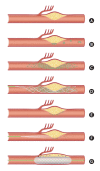
Illustrations of ICAS-O in terms of stroke pathomechanism and ERT strategy. (A-C) Pathomechanism of cerebral infarction on ICAS-O. Border-zone and scattered infarctions can occur from some microemboli, from in situ thrombosis in ICAS lesion. Perforator infarctions can also occur from the propagation of the thrombosis. (D) Stent retrieval for ICAS-O. Routine first-line thrombectomy can effectively eliminate the major portion of in situ thrombi. (E and F) Endothelial cells are still inflamed and may cause reocclusion. Glycoprotein IIb/IIIa inhibitor can stabilize the irritable endothelium. (G) The location of nearby important perforators should be cautiously evaluated when angioplasty and/or stenting are considered. This procedure can block the perforators, thereby aggravating neurological deficits. ICAS-O, intracranial atherosclerosis-related occlusion; ERT, endovascular revascularization treatment.
Figure 3.
Figure 3.
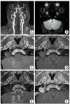
An embolism-related occlusion (EMB-O) case with vessel wall MRI taken on the third day after stroke onset. A 72-year-old man had an acute infarction in the posterior inferior cerebellar artery territory, due to right vertebral artery occlusion, but did not undertake endovascular revascularization treatment (ERT). His stroke etiology was cardioembolism by thorough evaluations. (A) Contrast-enhanced MR angiography shows an occlusion in the right vertebral artery. (B) Gradient echo images taken on admission, included in the baseline routine MRI, show a susceptibility vessel sign. Proton density-weighted (C) and T1-weighted (D) imaging does not show a specific finding but an occlusion is suggested since a signal void, as seen in normal arterial lumen, is absent. (E) T2-weighted imaging reveals an intact vessel wall but the lumen shows slightly higher signal intensity compared to a normal contralateral vertebral artery. (F) Thrombus in the occluded vessel appears to be stained by contrast and shows high signal intensity. The blue arrow indicates the vertebral artery occluded due to an embolism. The yellow arrow indicates the contralateral normal vertebral artery.

References

    1. Berkhemer OA, Fransen PS, Beumer D, van den Berg LA, Lingsma HF, Yoo AJ, et al. A randomized trial of intraarterial treatment for acute ischemic stroke. N Engl J Med. 2015;372:11–20. - PubMed
    1. Goyal M, Demchuk AM, Menon BK, Eesa M, Rempel JL, Thornton J, et al. Randomized assessment of rapid endovascular treatment of ischemic stroke. N Engl J Med. 2015;372:1019–1030. - PubMed
    1. Saver JL, Goyal M, Bonafe A, Diener HC, Levy EI, Pereira VM, et al. Stent-retriever thrombectomy after intravenous t-PA vs. t-PA alone in stroke. N Engl J Med. 2015;372:2285–2295. - PubMed
    1. Campbell BC, Mitchell PJ, Kleinig TJ, Dewey HM, Churilov L, Yassi N, et al. Endovascular therapy for ischemic stroke with perfusion-imaging selection. N Engl J Med. 2015;372:1009–1018. - PubMed
    1. Jovin TG, Chamorro A, Cobo E, de Miquel MA, Molina CA, Rovira A, et al. Thrombectomy within 8 hours after symptom onset in ischemic stroke. N Engl J Med. 2015;372:2296–2306. - PubMed