. 2017 Jun 7;7(1):3012.
doi: 10.1038/s41598-017-02533-2.

SNX10 gene mutation leading to osteopetrosis with dysfunctional osteoclasts

Affiliations

SNX10 gene mutation leading to osteopetrosis with dysfunctional osteoclasts

Eva-Lena Stattin et al. Sci Rep. .

Abstract

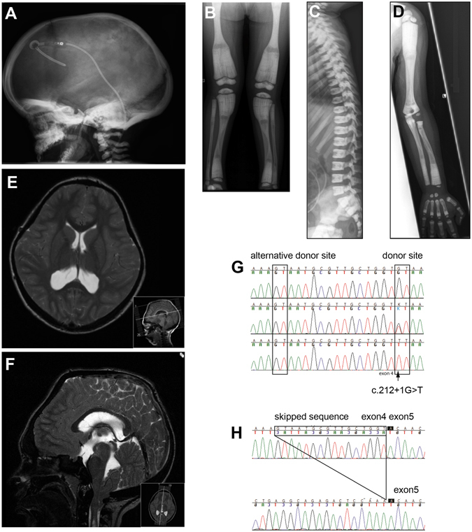
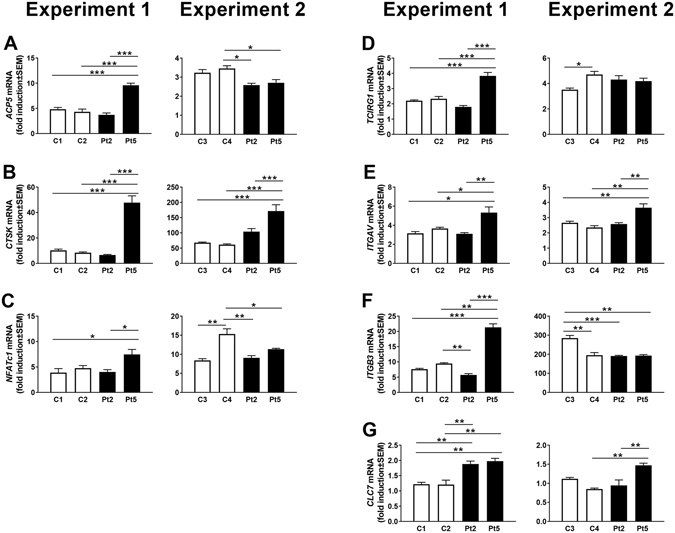
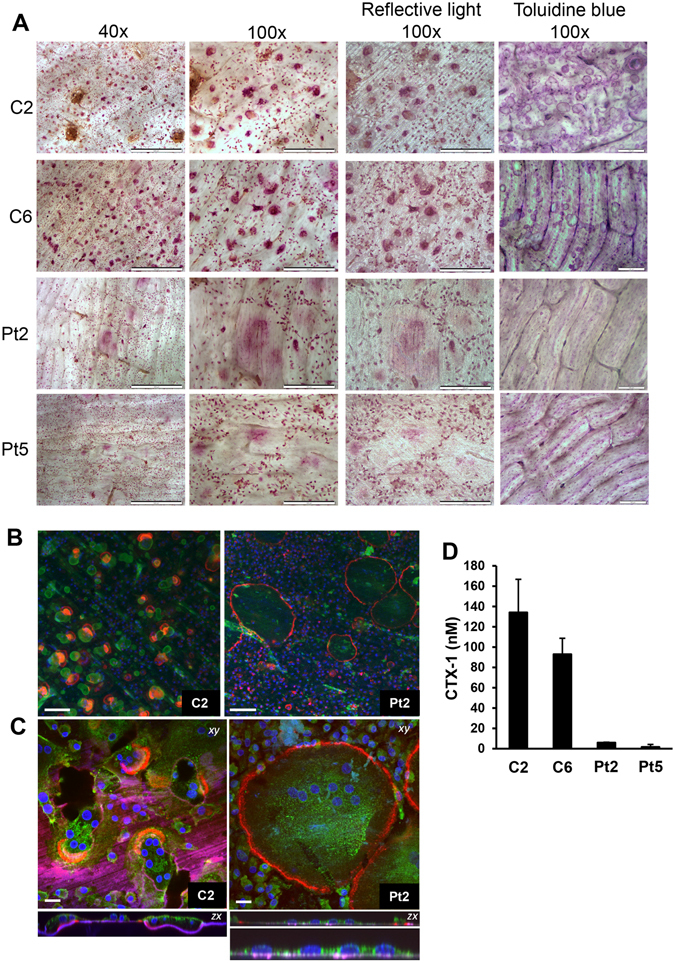
Autosomal recessive osteopetrosis (ARO) is a heterogeneous disorder, characterized by defective osteoclastic resorption of bone that results in increased bone density. We have studied nine individuals with an intermediate form of ARO, from the county of Västerbotten in Northern Sweden. All afflicted individuals had an onset in early infancy with optic atrophy, and in four patients anemia was present at diagnosis. Tonsillar herniation, foramen magnum stenosis, and severe osteomyelitis of the jaw were common clinical features. Whole exome sequencing, verified by Sanger sequencing, identified a splice site mutation c.212 + 1 G > T in the SNX10 gene encoding sorting nexin 10. Sequence analysis of the SNX10 transcript in patients revealed activation of a cryptic splice site in intron 4 resulting in a frame shift and a premature stop (p.S66Nfs * 15). Haplotype analysis showed that all cases originated from a single mutational event, and the age of the mutation was estimated to be approximately 950 years. Functional analysis of osteoclast progenitors isolated from peripheral blood of patients revealed that stimulation with receptor activator of nuclear factor kappa-B ligand (RANKL) resulted in a robust formation of large, multinucleated osteoclasts which generated sealing zones; however these osteoclasts exhibited defective ruffled borders and were unable to resorb bone in vitro.


Conflict of interest statement

The authors declare that they have no competing interests.

Figures

Figure 1
Figure 1
A Radiographs of subjects with the Västerbotten form of intermediate osteopetrosis, and sequencing of the SNX10 gene. (AD) Skeletal radiographs of a girl at the age of three years (Pt9), showing characteristic features of intermediate osteopetrosis. (A) Skull: Increased bone density of the skull base, frontal bossing and VP-shunt (B) AP view of the legs: “Erlenmeyer flask” shape of the femoral diaphysis. Longitudinal and transverse bands of lesser density in the metaphyses. Bone-within-bone pattern in the epiphyses in the knee. (C) Lateral view of the spine: Very dens endplates of the vertebral bodies, so called “sandwich vertebrae”. The ribs (seen in part) are broad and dens. (D) AP view of the left arm: No fractures or sign of rickets, “bone-within-bone” pattern of the phalanges. (E,F) Magnetic resonance imaging of the brain, and spine in an 11-year-old boy. MRI shows cerebellar tonsillar descent through the foramen magnum, a foramen magnum narrowing, and signs of brain stem compression with resulting syringo-hydromyelia. (G,H) Consequence of the c.212 + 1 G > T variant on (G) genomic level and (H) transcript level. (G) Sequencing of DNA from a control (top), a heterozygous carrier (middle) and a homozygous patient (bottom) for the c.212 + 1 G > T variant. The variant changes the donor splice site at the 5′ end of intron 4 from GT to TT. This leads to a predicted use of an alternative donor splice site 16 nucleotides upstream of intron 4. (H) Sequencing of cDNA from a control (top) and a patient (bottom) confirms the use of an alternative splice site and the skipping of 16 bp leading to a frame shift and a premature stop codon (p.S66Nfs * 15) at the protein level.
Figure 2
Figure 2
Osteoclast differentiation of CD14 + monocytes from peripheral blood. Photographs of TRAP staining (A) and quantification of surface area (B) of CD14 + cells cultured in M-CSF (M) or M-CSF and RANKL (M/RL) for four days (C1, C2, Pt2, Pt5) or five days (C1, lower photos, and Pt1). Surfaces are presented as mean ± SEM. (C) Actin ring staining of cells cultured in M/RL for four days. Scale bars: 100 μm. *** P ≤ 0.001.
Figure 3
Figure 3
Gene expression in osteoclasts from patients (Pt2, Pt5), and controls (C1, C2, C3, C4). Fold induction of gene expression between cells treated with M-CSF (M) with and without RANKL (RL) for three days. All values are given as mean ± SEM (n = 4). *P ≤ 0.05, **P ≤ 0.005, ***P ≤ 0.001 between indicated groups.
Figure 4
Figure 4
Patient-derived osteoclasts form actin rings but are unable to resorb bone. (A) TRAP staining of CD14 + cells cultured in M/RL on bone for eight days. Scale bar 40× panel: 500 μm, scale bar 100× light microscope and reflective light panel: 200 μm, scale bar 100× toluidine blue panel: 100 μm. (B,C) Osteoclasts were analyzed by confocal microscopy; (B) stained with TRITC-phalloidin (red), DAPI (blue) and the bone surface using a fluorescent bisphosphonate (AF-ALN; green). Resorption pits stain brightly with AF-ALN and can be seen in the control only. Scale bar = 100 μm; (C) stained with TRITC-phalloidin (red), DAPI (blue) AF-ALN (magenta) and the cell membranes with wheat germ agglutinin (green). AF-ALN omitted from upper panel of Pt2 for clarity. Black patches in C2 xy image indicates resorption pits. Bottom panel of Pt2 is a zoom of the zx section illustrating lack of resorption. Scale bar = 20 μm; (D) CTX-1 release in media during day 6 to 8 of culture in M/RL. C1, C6 = controls, Pt2, Pt5 = patients (n = 4–5).
Figure 5
Figure 5
Patient-derived osteoclasts formed defective ruffled borders. (A) Phalloidin staining for acting rings (left) and lectin staining for ruffled border (middle) of CD14 + cells cultured in M/RL on bone for 11 days. (BE)-Representative TEM micrographs of control C2 (B,C) and patient Pt2 (D,E) -derived osteoclasts show extensive ruffled border (grade 3) and resorption in the control, compared to lack of ruffled border (grade 0) and resorption in the patient. Patient osteoclasts formed sealing zones (E). N = nucleus, SZ = sealing zone, RB = ruffled border. Scale bars in left panels = 2 μm. Scale bars in right panels = 500 nm. (F) The percentage of osteoclasts categorized into each ruffled border grade (control, n = 38; patient, n = 34).
Figure 6
Figure 6
Expression of SNX10 mRNA in cells treated with M or M/RL for three days, analyzed with an assay covering the exon 1–2 (A) and exon 4–5 (B) junction. Values are given as mean ± SD (n = 2–4). ***P ≤ 0.001 between M and M/RL treated cells.

Similar articles

Cited by

References

    1. Frattini A, et al. Defects in TCIRG1 subunit of the vacuolar proton pump are responsible for a subset of human autosomal recessive osteopetrosis. Nature genetics. 2000;25:343–346. doi: 10.1038/77131. - DOI - PubMed
    1. Kornak U, et al. Loss of the ClC-7 chloride channel leads to osteopetrosis in mice and man. Cell. 2001;104:205–215. doi: 10.1016/S0092-8674(01)00206-9. - DOI - PubMed
    1. Sobacchi C, et al. The mutational spectrum of human malignant autosomal recessive osteopetrosis. Human molecular genetics. 2001;10:1767–1773. doi: 10.1093/hmg/10.17.1767. - DOI - PubMed
    1. Sobacchi C, Schulz A, Coxon FP, Villa A, Helfrich MH. Osteopetrosis: genetics, treatment and new insights into osteoclast function. Nature reviews. Endocrinology. 2013;9:522–536. doi: 10.1038/nrendo.2013.137. - DOI - PubMed
    1. Villa A, Guerrini MM, Cassani B, Pangrazio A, Sobacchi C. Infantile malignant, autosomal recessive osteopetrosis: the rich and the poor. Calcified tissue international. 2009;84:1–12. doi: 10.1007/s00223-008-9196-4. - DOI - PubMed

Publication types