Review
. 2017 Jul;18(7):389-403.
doi: 10.1038/nrn.2017.56. Epub 2017 Jun 8.

Functional consequences of neuropeptide and small-molecule co-transmission

Affiliations
Review

Functional consequences of neuropeptide and small-molecule co-transmission

Michael P Nusbaum et al. Nat Rev Neurosci. 2017 Jul.

Abstract

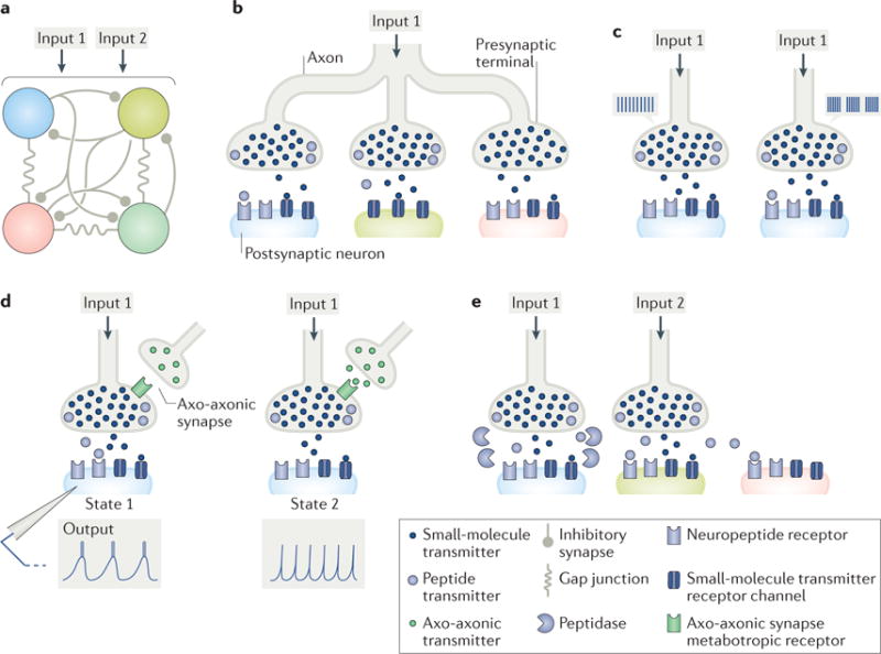
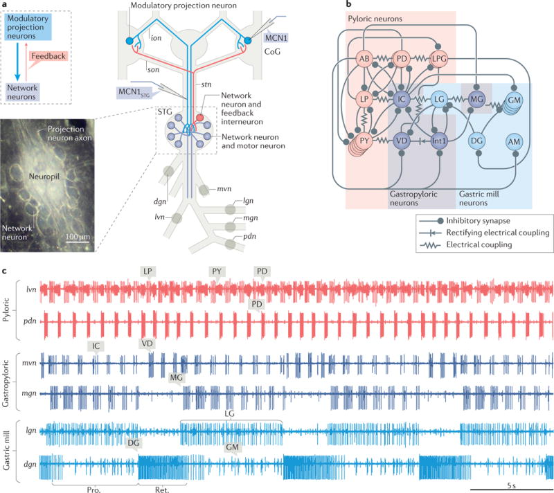
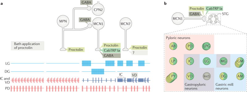
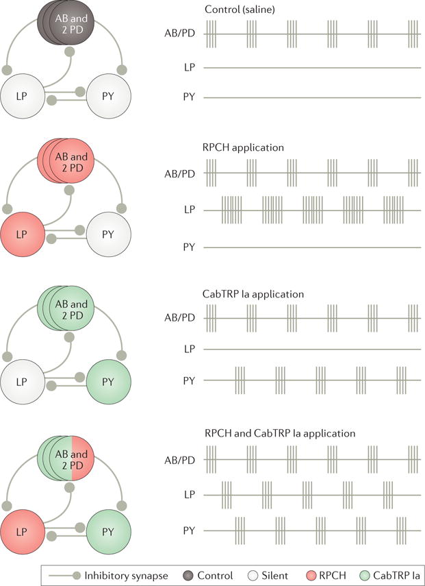
Colocalization of small-molecule and neuropeptide transmitters is common throughout the nervous system of all animals. The resulting co-transmission, which provides conjoint ionotropic ('classical') and metabotropic ('modulatory') actions, includes neuropeptide- specific aspects that are qualitatively different from those that result from metabotropic actions of small-molecule transmitter release. Here, we focus on the flexibility afforded to microcircuits by such co-transmission, using examples from various nervous systems. Insights from such studies indicate that co-transmission mediated even by a single neuron can configure microcircuit activity via an array of contributing mechanisms, operating on multiple timescales, to enhance both behavioural flexibility and robustness.


Conflict of interest statement

Competing interests statement

The authors declare no competing interests.

Figures

Figure 1
Figure 1. Co-transmission of small molecules and neuropeptides provides many degrees of freedom to microcircuit output
a| A schematic four-neuron microcircuit, interconnected by inhibitory synapses and electrical coupling, receiving input from two small-molecule–neuropeptide co-transmitting projection neurons (Input 1, Input 2) is shown. b | Co-transmission enables a single presynaptic neuron to have distinct actions on different postsynaptic neurons. Input 1 influences the blue circuit neuron via both co-transmitters, and the green and pink circuit neurons via only its small-molecule co-transmitter, albeit through different mechanisms (green neuron: no peptide receptors (postsynaptic mechanism); pink neuron: no peptide released nearby (presynaptic mechanism)). c | Co-transmitters can have distinct activity thresholds for their release. For example, Input 1 influences the blue circuit neuron by releasing only its small-molecule transmitter when firing at a low, tonic frequency (left), but it influences this circuit neuron by releasing both co-transmitters when firing in a rhythmic bursting pattern (right). d | Co-transmission is state dependent. Here, the state change is presynaptic, resulting from an axo-axonic synapse (green transmitter). When the axo-axonic synapse is not active (state 1), Input 1 co-releases both transmitters, and the blue circuit neuron responds with a rhythmic bursting pattern. When the axo-axonic synapse is active (state 2), peptide release is inhibited, and the blue circuit neuron responds with a tonic firing pattern. e | The diffusion distance of a neuronally released peptide can be regulated by the location of extracellular peptidases. Here, extracellular peptidases limit receptor access of Input 1-released peptide to the blue circuit neuron but do not limit Input 2-released peptide. Consequently, the green and pink circuit neurons are unlikely to be influenced by Input 1-released peptide, whereas both would be responsive to Input 2-released peptide, in addition to the green circuit neuron responding to the co-released small-molecule transmitter from Input 2. The peptidase activity near the blue circuit neuron would limit or possibly prevent its response to Input 2-released peptide.
Figure 2
Figure 2. The crab Cancer borealis stomatogastric nervous system
a| A schematic of the isolated stomatogastric nervous system of the crab Cancer borealis is shown. The inset shows a whole-mount image of the desheathed stomatogastric ganglion (STG) under dark-field illumination (anterior, top; posterior, bottom). As it is evident in both the schematic and the inset, the 26 neuronal somata form a single layer surrounding the neuropil. Circles on nerves indicate recording sites for traces shown in part c. b | A schematic of the gastric mill and pyloric circuit is shown. The arrangement of neurons in the schematic represents the relative timing of activity for each neuron during the gastric mill and pyloric rhythms. Specifically, the neurons that exhibit pyloric rhythm-timed activity (‘pyloric neurons’ and ‘gastropyloric neurons’) are displayed such that the top-row neurons are co-active, followed by the middle-row neurons and then the bottom-row neurons, after which the top-row neurons are again active. The neurons that exhibit gastric mill rhythm-timed activity (‘gastric mill neurons’ and ‘gastropyloric neurons’) are displayed such that the top-row neurons are co-active and burst in alternation with the bottom-row neurons. As shown, there are eight gastric mill circuit neuron types, one of which is present as four apparently equivalent copies (GM neurons). All eight neuron types contribute to gastric mill pattern generation, whereas only two (LG and Int1) are also rhythm generator neurons,,. There are seven pyloric circuit neuron types, including the rhythm generator (‘pacemaker’) group AB/PD/LPG. Three of these neuron types are present as multiple, apparently equivalent copies (PD: 2; LPG: 2; PY: 5). c | Simultaneous extracellular nerve recordings of the gastric mill and pyloric rhythms during tonic stimulation of the modulatory projection neuron modulatory commissural neuron 1 (MCN1) are shown. The pyloric rhythm exhibits a rhythmically repeating triphasic pattern (for example, lateral ventricular nerve (lvn): PD, LP, PY) that is continuously active, in vivo and in vitro, with a cycle period of ~1 s. The gastric mill rhythm (cycle period ~10–20 s) is silent except when driven by modulatory neurons (for example, MCN1), which themselves require activation in vivo and in vitro. It is a rhythmically repeating biphasic pattern, consisting of teeth protraction (Pro.) and teeth retraction (Ret.), which drives the motor response (chewing). Note that some neurons exhibit activity patterns time-locked to both rhythms (gastropyloric neurons). CoG, commissural ganglion; dgn, dorsal gastric nerve; ion, inferior oesophageal nerve; lgn, lateral gastric nerve; mgn, medial gastric nerve; mvn, medial ventricular nerve; pdn, pyloric dilator nerve; son, superior oesophageal nerve; stn, stomatogastric nerve. Part b is adapted with permission from REF., Macmillan Publishers Limited. STG photo courtesy of Marie Suver, New York University, USA, and Wolfgang Stein, Illinois State University, USA.
Figure 3
Figure 3. The microcircuit response to peptidergic neuron activity is not necessarily mimicked by bath application of that neuropeptide
a| This part shows schematic extracellular recordings of identified neurons in the crab Cancer borealis stomatogastric ganglion (STG), which are active during the gastric mill rhythm (LG and DG neurons), pyloric rhythm (PD neuron) or both rhythms (IC and VD neurons). In the isolated crab STG, bath-applied proctolin (far left set of responses) selectively excites the pyloric rhythm,. This action mimics the response to activation of only one (modulatory proctolin neuron (MPN)) of the three proctolinergic projection neurons that innervate the STG (MPN, modulatory commissural neuron 1 (MCN1) and MCN7), even though MPN also contains a small-molecule co-transmitter (GABA),. As indicated, MPN also inhibits two projection neurons (MCN1 and commissural projection neuron 2 (CPN2)) by releasing GABA from a separate axon projecting to a separate location (commissural ganglion (CoG)),. The other two proctolinergic projection neurons (MCN1 and MCN7) also influence STG microcircuit activity but elicit activity patterns from the circuit neurons that are distinct from proctolin bath application,. MCN1-released C. borealis tachykinin-related peptide Ia (CabTRP Ia) and GABA are pivotal for MCN1 activation of the gastric mill rhythm, whereas its release of CabTRP Ia and proctolin dominates its excitation of the pyloric rhythm (see part b). The MCN7 actions on these rhythms result partly from proctolin and probably also from one or more yet-to-be-identified co-transmitters (indicated by ‘?’). In the figure, pyloric rhythm activity is shown in red; gastric mill rhythm activity is shown in blue; gastropyloric activity is shown in purple. b | In the crab STG, MCN1 innervates all pyloric, gastropyloric and gastric mill neurons. The figure shows a representation of responsiveness of each STG circuit neuron to the MCN1-released co-transmitters proctolin (light green), CabTRP Ia (dark green) and GABA (dark grey),. Examples of convergent peptide co-transmitter action (proctolin and CabTRP Ia), selective peptide co-transmitter action (CabTRP Ia) and selective GABA action are shown. In some cases, the STG neuron only responds to the indicated co-transmitter (or co-transmitters) (for example, Int1). In other cases, the STG neuron does respond to an additional co-transmitter but not when it is released from MCN1 (for example, LG responds to applied GABA but not GABA released from MCN1). No information is available regarding whether these co-transmitters are colocalized to all MCN1 terminals or are localized to separate terminals for their release. Part a is adapted with permission from REF., Elsevier.
Figure 4
Figure 4. Peptide co-transmitters can have complementary actions on microcircuit output
This figure shows the schematic recordings of the core pyloric circuit neurons in the isolated stomatogastric ganglion (STG) of the lobster Homarus americanus under control conditions (saline) and during bath application of the peptide co-transmitters red pigment-concentrating hormone (RPCH) or Cancer borealis tachykinin-related peptide Ia (CabTRP Ia), or both together. Before STG isolation, the complete pyloric rhythm is expressed, including rhythmic sequential bursting of circuit neurons AB/PD, LP and PY (not shown). Under control conditions in the isolated STG, only the pyloric pacemaker ensemble (AB and both PDs) remains rhythmically active. Applying RPCH alone recruits the LP neuron to resume pyloric-timed bursting, whereas applying CabTRP Ia alone recruits pyloric-timed bursting in the PY neuron. Co-applying both peptides reactivates the complete pyloric rhythm. Adapted with permission of Society for Neuroscience from: Colocalized neuropeptides activate a central pattern generator by acting on different circuit targets, Thirumalai V. & Marder E., J. Neurosci. 22, 1874–1882, 2002; permission conveyed through Copyright Clearance Center, Inc.
Figure 5
Figure 5. The response of a microcircuit to co-transmission can be sculpted by feedback to the co-transmitting neuron
a| Modulatory commissural neuron 1 (MCN1) synaptic interactions with the gastric mill microcircuit are shown. The core rhythm generator includes the synaptic interactions between and intrinsic properties of MCN1, LG and Int1. These three neurons are necessary and sufficient to generate the gastric mill rhythm. The pyloric pacemaker interneuron AB regulates the gastric mill rhythm generator via its inhibitory synapse onto Int1. However, AB is not necessary for rhythm generation; the rhythm slows but continues when AB activity is eliminated,. The motor neurons shown in grey are not necessary for rhythm generation, but some contribute to pattern generation via their intra-circuit synapses, and all contribute to movement via their synapses onto specific gastric mill muscles. The top row shows gastric mill protractor neurons (that is, motor neurons where their activation causes the teeth to move towards each other); the bottom row shows retractor neurons (that is, motor neurons, the activation of which causes the teeth to move away from one another and that are co-active with Int1 neuron); AB and PD are pyloric pacemaker neurons. All neurons are motor neurons except Int1 and AB, which are interneurons that project to the commissural ganglion (CoG). MCN1 excitation of Int1 is ionotropic; all other MCN1 co-transmitter actions are metabotropic. b | Tonic MCN1 stimulation drives the gastric mill rhythm (LG and Int1) and speeds up the pyloric rhythm (AB). MCN1 activation of the rhythm generator neurons LG and Int1 establishes the rhythmic alternating bursting pattern (LG bursting during the teeth protraction (Pro.) phase; Int1 bursting during the teeth retraction (Ret.) phase) that is then imposed on all gastric mill neurons. c | The gastric mill rhythm generator circuit during Ret. and Pro. phases of the MCN1-gastric mill rhythm, is shown. During Ret. (left), MCN1 activity causes co-transmitter release, which drives Int1 activity, via ionotropic (i) excitation and provides a slow build-up of metabotropic (m) excitation that eventually enables LG to escape from Int1 inhibition and fire a burst of action potentials. The onset of a LG burst triggers the switch to the Pro. phase (right), during which LG is active and inhibits Int1. During Pro., LG activity also provides ionotropic inhibition to the stomatogastric ganglion (STG) terminals of MCN1 that prevents further co-transmitter release from MCN1 but enables MCN1 activity to sustain LG activity via an electrical synapse until the slowly decaying metabotropic excitation in LG falls below a critical level (see part d). In the figure, active neurons and synapses are shown in blue; silent neurons and synapses are shown in grey; the slanted double hashmarks indicate the abbreviated MCN1 axon. d | This part shows the output of a computational model showing the MCN1 Cancer borealis tachykinin-related peptide Ia (CabTRP Ia)-activated conductance GMI–MCN1 in the LG neuron waxing and waning during the gastric mill Ret. and Pro. phases, respectively (lower VLG trace). GMI–MCN1 increases during Ret. owing to continual CabTRP Ia release from MCN1, whereas it decays during Pro. owing to LG inhibition of the MCN1 terminals in the STG (see part c). The peak conductance occurs at the LG burst onset threshold, whereas the steep drop in conductance at the end of the LG burst occurs at the LG burst offset threshold. Part a is adapted with permission of Society for Neuroscience from: Convergent rhythm generation from divergent cellular mechanisms, Rodriguez J. C., Blitz D. M. & Nusbaum M. P., J. Neurosci. 33, 18047–18064, 2013; permission conveyed through Copyright Clearance Center, Inc. Part b is adapted with permission of Society for Neuroscience from: Coordination of fast and slow rhythmic neuronal circuits, Bartos M., Manor Y., Nadim F., Marder E. & Nusbaum M. P., J. Neurosci. 19, 6650–6660, 1999; permission conveyed through Copyright Clearance Center, Inc. Part c is adapted with permission from REF., Macmillan Publishers Limited. Part d is adapted with permission of Society for Neuroscience from: Parallel regulation of a modulator-activated current via distinct dynamics underlies comodulation of motor circuit output, DeLong N. D., Kirby M. S., Blitz D. M. & Nusbaum M. P., J. Neurosci. 29, 12355–12367, 2009; permission conveyed through Copyright Clearance Center, Inc.
Figure 6
Figure 6. The muscle stretch-sensitive GPR neuron causes a state-dependent prolongation of the gastric mill retractor phase by selectively inhibiting CabTRP Ia release from MCN1
Aa| The schematic recordings show that stimulating the gastropyloric receptor neurons (GPR) during an ongoing modulatory commissural neuron 1 (MCN1)-stimulated gastric mill rhythm delays LG firing and selectively prolongs the gastric mill retractor phase,,,. Ab | The schematic circuit diagram depicts that, during the MCN1-gastric mill rhythm, GPR stimulation selectively inhibits Cancer borealis tachykinin-related peptide Ia (CabTRP Ia) release from MCN1 (represented by the smaller MCN1 terminal onto LG, as well as the placement of the GPR synapse) using only one of its co-transmitters (5-hydroxytryptamine (5-HT)),,,. By reducing CabTRP Ia release from MCN1, GPR slows the build-up of modulator-activated inward current (IMI) in LG, thereby delaying the next LG burst onset. The grey-coloured GPR synapses are too weak to influence the rhythm relative to the other synaptic events occurring during the MCN1-gastric mill rhythm,,,. In the figure, the MCN1 co-transmitter separation is only for schematic presentation, as it is not known whether there is a spatial separation of the MCN1 co-transmitters to different terminals. Ba | The schematic recordings show that the prolongation of the gastric mill retractor phase caused by GPR stimulation during the MCN1-stimulated gastric mill rhythm in the presence of normal saline (part Aa) is suppressed by the peptide hormone crustacean cardioactive peptide (CCAP). CCAP also strengthens and slightly prolongs the protractor phase (LG burst) without altering retractor phase duration,. Bb | CCAP and MCN1-released CabTRP Ia bind to separate G protein-coupled receptors (GPCRs) on the LG neuron, but nevertheless each activates IMI current in LG,,,. GPR is silent (grey). Bc | The amount of IMI activation is not reduced when GPR is active (blue) and inhibiting CabTRP Ia release from MCN1, owing to the parallel IMI activation by CCAP,,,. ACh, acetylcholine; AST, A type allatostatin; CoG, commissural ganglion; STG, stomatogastric ganglion. Part Ab is adapted with permission from REF., APS. Part Bb and part c are adapted with permission of Society for Neuroscience from: Hormonal modulation of sensorimotor integration, DeLong N. D. & Nusbaum M. P., J. Neurosci. 30, 2418–2427, 2010; permission conveyed through Copyright Clearance Center, Inc.

Similar articles

Cited by

References

    1. Lingueglia E, Champigny G, Lazdunski M, Barbry P. Cloning of the amiloride-sensitive FMRFamide peptide-gated sodium channel. Nature. 1995;378:730–733. - PubMed
    1. Durrnagel S, et al. Three homologous subunits form a high affinity peptide-gated ion channel in Hydra. J Biol Chem. 2010;285:11958–11965. - PMC - PubMed
    1. Grunder S, Assmann M. Peptide-gated ion channels and the simple nervous system of Hydra. J Exp Biol. 2015;218:551–561. - PubMed
    1. Trudeau LE, et al. The multilingual nature of dopamine neurons. Prog Brain Res. 2014;211:141–164. - PMC - PubMed
    1. Tritsch NX, Granger AJ, Sabatini BL. Mechanisms and functions of GABA co-release. Nat Rev Neurosci. 2016;17:139–145. - PMC - PubMed

Publication types

LinkOut - more resources