. 2016 Nov;28(11):2644-2658.
doi: 10.1002/elan.201600183. Epub 2016 Jun 7.

Multiplex Immunosensor Arrays for Electrochemical Detection of Cancer Biomarker Proteins

Affiliations

Multiplex Immunosensor Arrays for Electrochemical Detection of Cancer Biomarker Proteins

Bernard S Munge et al. Electroanalysis. 2016 Nov.

Abstract

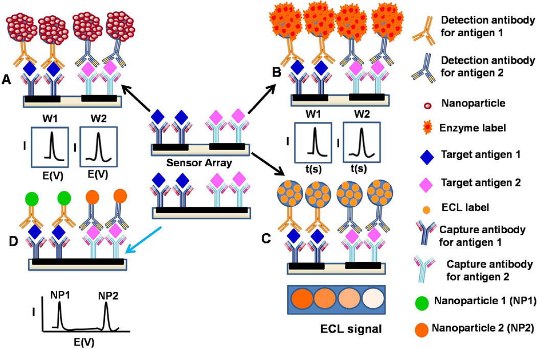
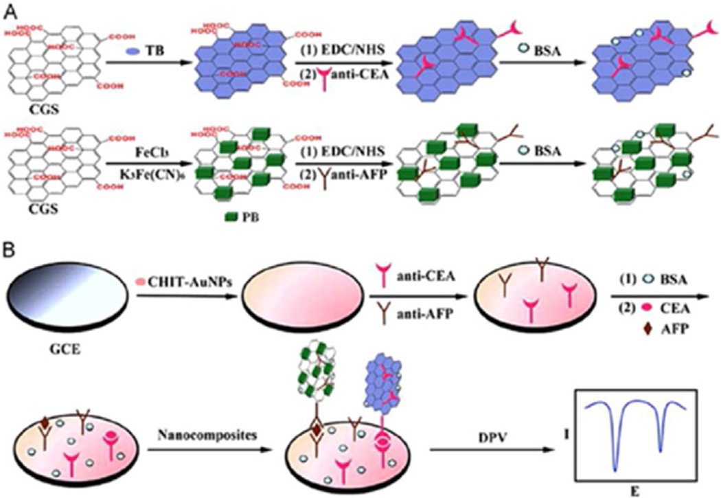
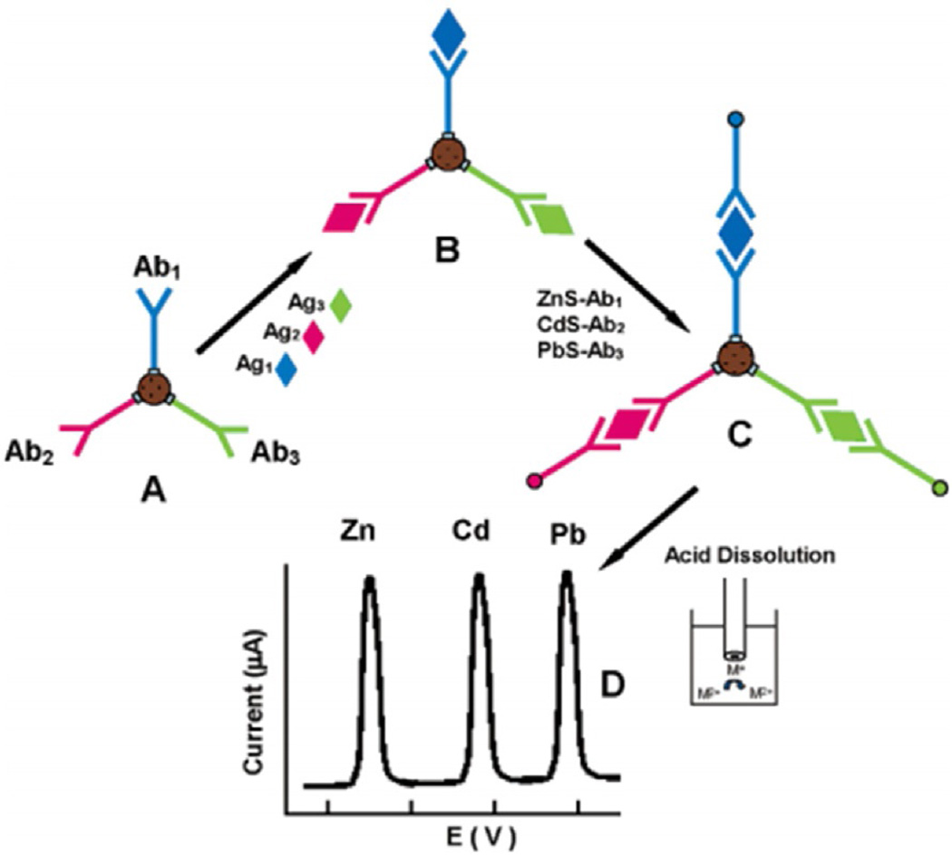
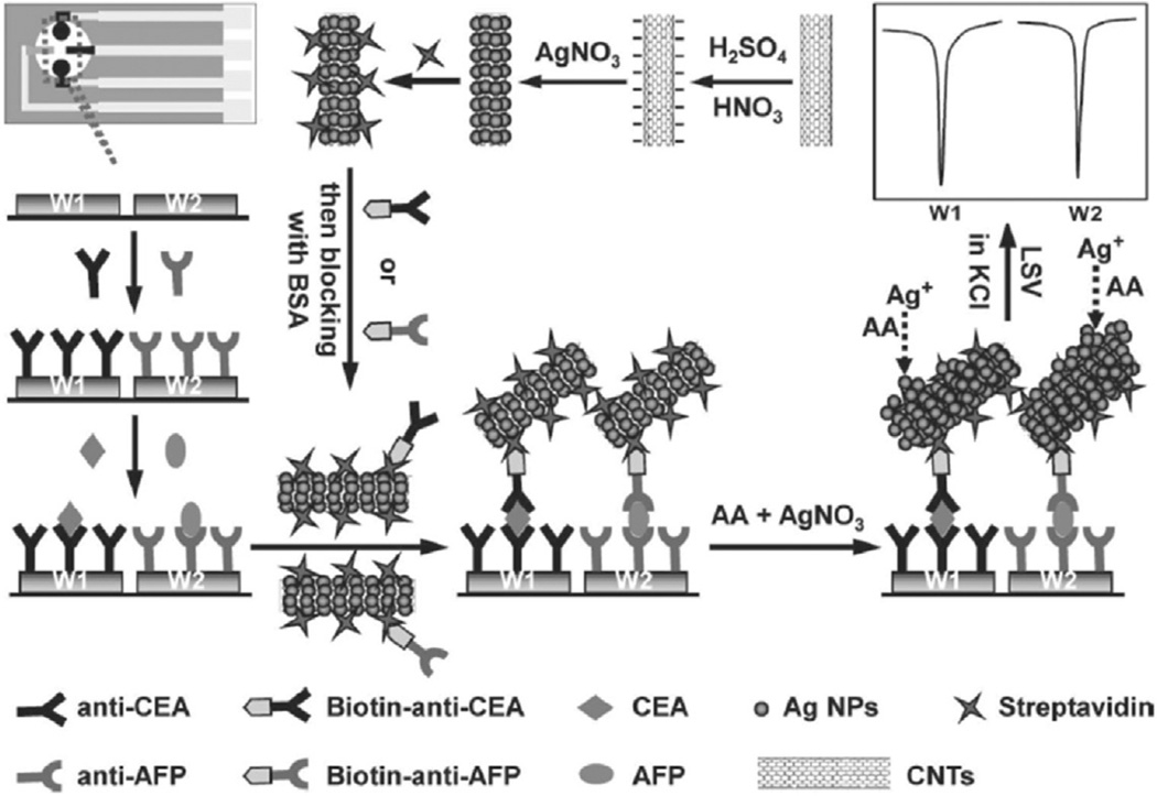
Measuring panels of protein biomarkers offer a new personalized approach to early cancer detection, disease monitoring and patients' response to therapy. Multiplex electrochemical methods are uniquely positioned to provide faster, more sensitive, point of care (POC) devices to detect protein biomarkers for clinical diagnosis. Nanomaterials-based electrochemical methods offer sensitivity needed for early cancer detection. This review discusses recent advances in multiplex electrochemical immunosensors for cancer diagnostics and disease monitoring. Different electrochemical strategies including enzyme-based immunoarrays, nanoparticle-based immunoarrays and electrochemiluminescence methods are discussed. Many of these methods have been integrated into microfluidic systems, but measurement of more than 2-4 protein markers in a small single serum sample is still a challenge. For POC applications, a simple, low cost method is required. Major challenges in multiplexed microfluidic immunoassays are reagent additions and washing steps that require creative engineering solutions. 3-D printed microfluidics and paper-based microfluidic devices are also explored.

Keywords: Cancer biomarker protein; Electrochemical Immunosensor; Electrochemiluminescence; Microfluidic; Multiplex.


Figures

Fig. 1
Fig. 1
Strategies for multiplex electrochemical detection of proteins using a multi-electrode array with separate multiple microwells modified with different capture antibodies for different target biomarker proteins in a sandwich type assay; (A) coupled to multi-nanoparticle labels for enhanced sensitivity, (B) using multi-enzyme labels for enhanced sensitivity (C) Electrochemiluminescence tag and (D) an alternative approach using a single electrode well e.g. GCE or Magnetic beads with different nanocrystal labels or redox probes for distinguishable electrochemical signals. Multi-electrode arrays can also be integrated to microfluidic systems to reduce sample volume, automate the assay process and increase throughput.
Fig. 2
Fig. 2
Illustration of SPCE array with multiple elements for multiplex electrochemical detection of proteins. The working electrode elements are modified with different capture antibodies for a sandwich type assay coupled to universal MWNT bioconjugates with multi-enzyme labels for enhanced sensitivity. Adapted with permission from ref. [55]. Copyright 2011 Elsevier.
Fig. 3
Fig. 3
Multiplex electrochemical detection using a single well glassy carbon electrode (GCE) modified with a mixture of capture antibodies for different target biomarker proteins. (A) preparation of biofunctional graphene tag containing 2 different redox probes, toluidine blue and Prussian blue for distinguishable signals, and (B) multiplexed electrochemical immunoassay protocol. Reprinted from ref. [61]. Copyright 2013 Elsevier.
Fig. 4
Fig. 4
Multiplex electrochemical detection based on Qdots for distinguishable signals. (A) Different capture antibodies attached to a magnetic bead, followed by (B) binding of the corresponding target antigens on the magnetic beads; (C) Binding of the Qdot-labeled secondary antibodies to form a sandwich type assay (D) acid dissolution of Qdots and subsequent electrochemical stripping detection. Adapted from ref. . Copyright 2004 American chemical society.
Fig. 5
Fig. 5
Schematic representation of sandwich type nanoparticle-based immunoarray fabricated on 2 separate microwells with different capture antibodies for different target biomarker detection. Schematic shows preparation of multi-nanoparticles trace tag, and detection strategy by linear-sweep stripping voltammetric analysis of AgNPs on the immunosensor surface without acid dissolution. Adapted from reference . Copyright 2011 Wiley.
Fig. 6
Fig. 6
Electrochemical proteins detected in serum by multi-electrode array integrated into microfluidic system featuring a capture and a detection channel made from PDMS encased in polymethylmethacrylate plastic (A). The electrochemical sensor array was positioned in a PDMS microfluidic channel containing a platinum (Pt) wire as the counter electrode and a silver/silver chloride (Ag/ AgCl) wire reference electrode. Amperometry signal was generated upon incubation with Ab2-MB-HRP-analytes in the measurement chamber for 20 min, then injecting a mixture of H2O2 and HQ: (B) duplicate responses in simultaneous array measurements on a standard mixture of 10 fg mL−1 IL-6, 15 fg mL−1 IL-8, 25 fg mL−1 VEG, and 60 fg mL−1 VEGFC illustrating reproducibility, (C) responses to human VEGF in mixtures of biomarker proteins (peaks for VEGF were extracted from four-protein determinations and presented together), (D–G) corresponding Array calibration plots of standard mixtures in calf serum for IL-6 (D), IL-8 (E), VEGF (F), VEGF-C (G). Standard deviations correspond to 2 sensors each on three separate arrays (n = 6). Adapted from ref. . Copyright 2012 American Chemical Society.
Fig. 7
Fig. 7
Automated ECL Immunoarray: (A) microprocessor-controlled microfluidic immunoarray featuring a 30 microwell SWCNT modified detection array fed with sample/immunore-agents from a reagent cassette (red) using inexpensive micro-pumps. (B) automated immunoassay steps that are controlled by a microprocessor. ECL results (C–D) for a panel of four cancer biomarkers, IL-6, PF4, PSMA, PSA, at different target protein concentrations and E-H showing corresponding immunoassay calibration curves for IL-6, PF4, PSA, and PSMA. Adapted from ref. . Copyright 2015 American Chemical Society.

References

    1. Atkinson AJ, Colburn WA, DeGruttola VG, DeMets DL, Downing GJ, Hoth DF, Oates JA, G Peck C, Schooley RT, Spilker BA. Clin. Pharmacol. Ther. 2001;69:89–95. - PubMed
    1. Hawkridge AM, Muddiman DC. Annu. Rev. Anal. Chem. 2009;2:265–277. - PMC - PubMed
    1. Kulasingam V, Diamandis EP. Nat. Clin. Pract. Oncol. 2008;5:588–599. - PubMed
    1. Ludwig JA, Weinstein JN. Nat. Rev. Cancer. 2005;5:845–856. - PubMed
    1. Ferrari M. Nat. Rev. Cancer. 2005;5:161–171. - PubMed

LinkOut - more resources