

Malignant Peripheral Nerve Sheath Tumors State of the Science: Leveraging Clinical and Biological Insights into Effective Therapies
- PMID: 28592921
- PMCID: PMC5448069
- DOI: 10.1155/2017/7429697
Malignant Peripheral Nerve Sheath Tumors State of the Science: Leveraging Clinical and Biological Insights into Effective Therapies
Abstract
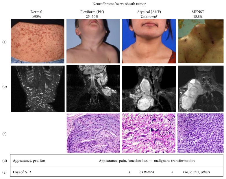
Malignant peripheral nerve sheath tumor (MPNST) is the leading cause of mortality in patients with neurofibromatosis type 1. In 2002, an MPNST consensus statement reviewed the current knowledge and provided guidance for the diagnosis and management of MPNST. Although the improvement in clinical outcome has not changed, substantial progress has been made in understanding the natural history and biology of MPNST through imaging and genomic advances since 2002. Genetically engineered mouse models that develop MPNST spontaneously have greatly facilitated preclinical evaluation of novel drugs for translation into clinical trials led by consortia efforts. Continued work in identifying alterations that contribute to the transformation, progression, and metastasis of MPNST coupled with longitudinal follow-up, biobanking, and data sharing is needed to develop prognostic biomarkers and effective prevention and therapeutic strategies for MPNST.
Figures
References
-
- Meany H., Widemann B. C., Ratner N. Neurofibromatosis Type 1. Berlin, Germany: Springer; 2012. Malignant peripheral nerve sheath tumors: prognostic and diagnostic markers and therapeutic targets; pp. 445–467.
Publication types
LinkOut - more resources
Full Text Sources
Other Literature Sources
Medical
Research Materials
