

Mechanisms Underlying the Essential Role of Mitochondrial Membrane Lipids in Yeast Chronological Aging
- PMID: 28593023
- PMCID: PMC5448074
- DOI: 10.1155/2017/2916985
Mechanisms Underlying the Essential Role of Mitochondrial Membrane Lipids in Yeast Chronological Aging
Abstract
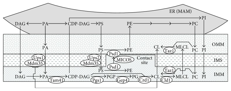
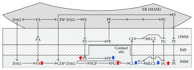
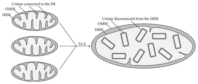
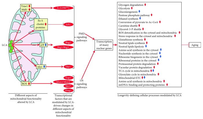
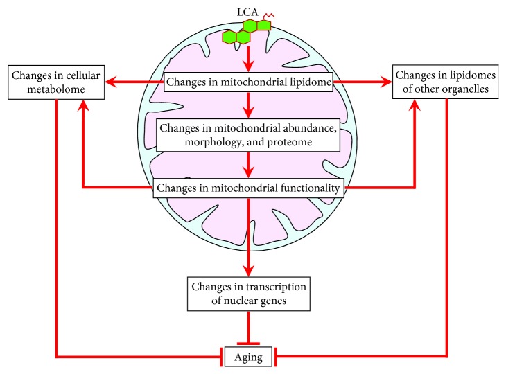
The functional state of mitochondria is vital to cellular and organismal aging in eukaryotes across phyla. Studies in the yeast Saccharomyces cerevisiae have provided evidence that age-related changes in some aspects of mitochondrial functionality can create certain molecular signals. These signals can then define the rate of cellular aging by altering unidirectional and bidirectional communications between mitochondria and other organelles. Several aspects of mitochondrial functionality are known to impact the replicative and/or chronological modes of yeast aging. They include mitochondrial electron transport, membrane potential, reactive oxygen species, and protein synthesis and proteostasis, as well as mitochondrial synthesis of iron-sulfur clusters, amino acids, and NADPH. Our recent findings have revealed that the composition of mitochondrial membrane lipids is one of the key aspects of mitochondrial functionality affecting yeast chronological aging. We demonstrated that exogenously added lithocholic bile acid can delay chronological aging in yeast because it elicits specific changes in mitochondrial membrane lipids. These changes allow mitochondria to operate as signaling platforms that delay yeast chronological aging by orchestrating an institution and maintenance of a distinct cellular pattern. In this review, we discuss molecular and cellular mechanisms underlying the essential role of mitochondrial membrane lipids in yeast chronological aging.
Figures
Similar articles
-
Lithocholic bile acid accumulated in yeast mitochondria orchestrates a development of an anti-aging cellular pattern by causing age-related changes in cellular proteome.Cell Cycle. 2015;14(11):1643-56. doi: 10.1080/15384101.2015.1026493. Cell Cycle. 2015. PMID: 25839782 Free PMC article.
-
Mitochondrial membrane lipidome defines yeast longevity.Aging (Albany NY). 2013 Jul;5(7):551-74. doi: 10.18632/aging.100578. Aging (Albany NY). 2013. PMID: 23924582 Free PMC article.
-
Specific changes in mitochondrial lipidome alter mitochondrial proteome and increase the geroprotective efficiency of lithocholic acid in chronologically aging yeast.Oncotarget. 2017 May 9;8(19):30672-30691. doi: 10.18632/oncotarget.16766. Oncotarget. 2017. PMID: 28410198 Free PMC article.
-
Communications between Mitochondria, the Nucleus, Vacuoles, Peroxisomes, the Endoplasmic Reticulum, the Plasma Membrane, Lipid Droplets, and the Cytosol during Yeast Chronological Aging.Front Genet. 2016 Sep 27;7:177. doi: 10.3389/fgene.2016.00177. eCollection 2016. Front Genet. 2016. PMID: 27729926 Free PMC article. Review.
-
Some Metabolites Act as Second Messengers in Yeast Chronological Aging.Int J Mol Sci. 2018 Mar 15;19(3):860. doi: 10.3390/ijms19030860. Int J Mol Sci. 2018. PMID: 29543708 Free PMC article. Review.
Cited by
-
ATF6 safeguards organelle homeostasis and cellular aging in human mesenchymal stem cells.Cell Discov. 2018 Jan 5;4:2. doi: 10.1038/s41421-017-0003-0. eCollection 2018. Cell Discov. 2018. PMID: 29423270 Free PMC article.
-
Mechanisms through which lithocholic acid delays yeast chronological aging under caloric restriction conditions.Oncotarget. 2018 Oct 9;9(79):34945-34971. doi: 10.18632/oncotarget.26188. eCollection 2018 Oct 9. Oncotarget. 2018. PMID: 30405886 Free PMC article.
-
The Dual Role of the Pervasive "Fattish" Tissue Remodeling With Age.Front Endocrinol (Lausanne). 2019 Feb 26;10:114. doi: 10.3389/fendo.2019.00114. eCollection 2019. Front Endocrinol (Lausanne). 2019. PMID: 30863366 Free PMC article. Review.
-
Mechanisms by which PE21, an extract from the white willow Salix alba, delays chronological aging in budding yeast.Oncotarget. 2019 Oct 8;10(56):5780-5816. doi: 10.18632/oncotarget.27209. eCollection 2019 Oct 8. Oncotarget. 2019. PMID: 31645900 Free PMC article.
-
Changes in Chromatin Organization Eradicate Cellular Stress Resilience to UVA/B Light and Induce Premature Aging.Cells. 2021 Jul 11;10(7):1755. doi: 10.3390/cells10071755. Cells. 2021. PMID: 34359924 Free PMC article.
References
Publication types
MeSH terms
Substances
LinkOut - more resources
Full Text Sources
Other Literature Sources
