. 2017 Apr 26;7(6):730-746.
doi: 10.1002/2211-5463.12219. eCollection 2017 Jun.

The neuronal and astrocytic protein SLC38A10 transports glutamine, glutamate, and aspartate, suggesting a role in neurotransmission

Affiliations

The neuronal and astrocytic protein SLC38A10 transports glutamine, glutamate, and aspartate, suggesting a role in neurotransmission

Sofie V Hellsten et al. FEBS Open Bio. .

Abstract

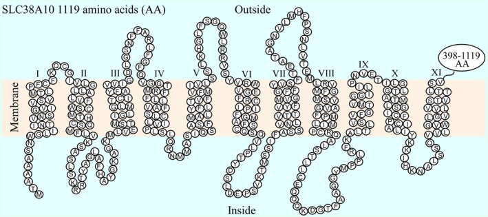
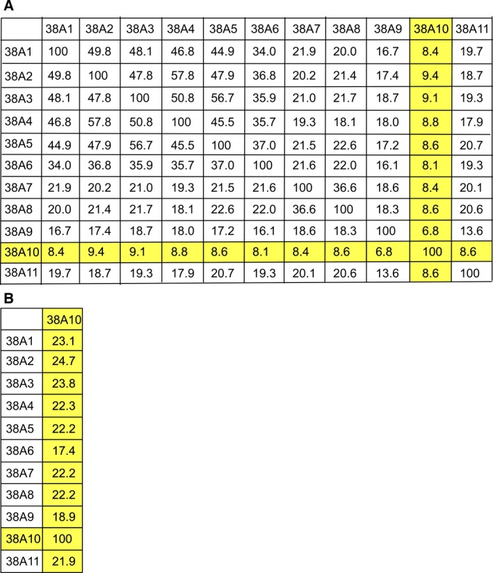
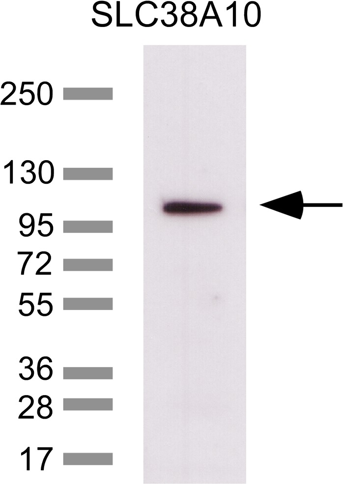
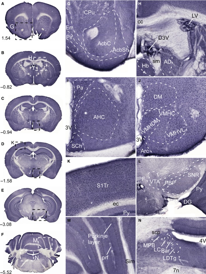
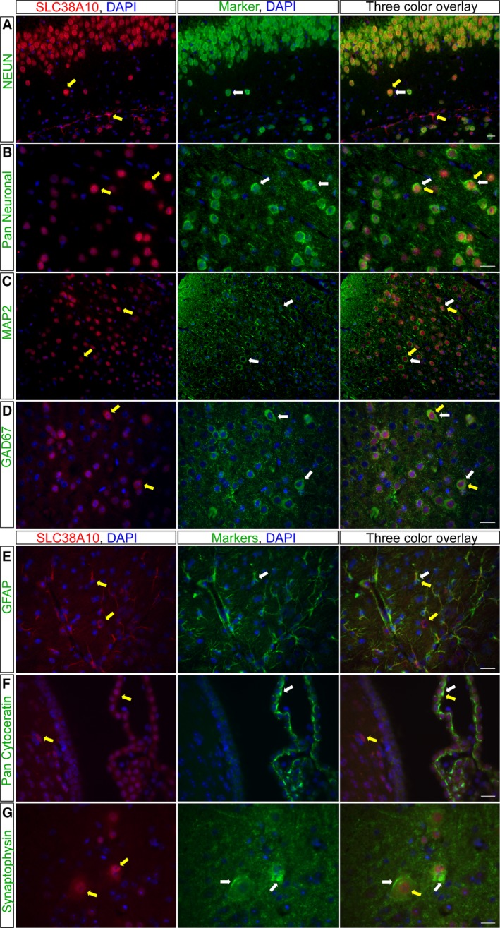
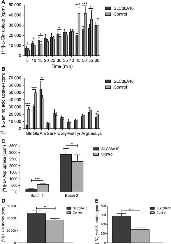
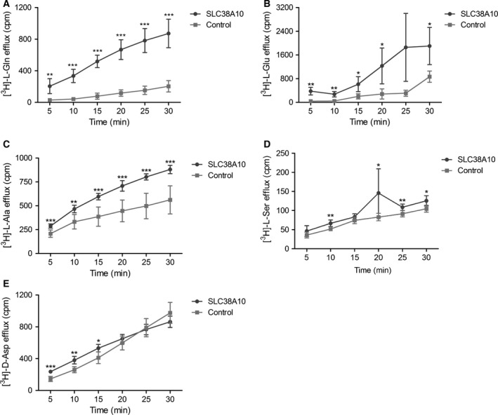
In brain cells, glutamine transporters are vital to monitor and control the levels of glutamate and GABA. There are 11 members of the SLC38 family of amino acid transporters of which eight have been functionally characterized. Here, we report the first histological and functional characterization of the previously orphan member, SLC38A10. We used pairwise global sequence alignments to determine the sequence identity between the SLC38 family members. SLC38A10 was found to share 20-25% transmembrane sequence identity with several family members, and was predicted to have 11 transmembrane helices. SLC38A10 immunostaining was abundant in mouse brain using a custom-made anti-SLC38A10 antibody and colocalization of SLC38A10 immunoreactivity with markers for neurons and astrocytes was detected. Using Xenopus laevis oocytes overexpressing SLC38A10, we show that SLC38A10 mediates bidirectional transport of l-glutamine, l-alanine, l-glutamate, and d-aspartate, and efflux of l-serine. This profile mostly resembles system A members of the SLC38 family. In conclusion, the bidirectional transport of glutamine, glutamate, and aspartate by SLC38A10, and the immunostaining detected in neurons and astrocytes, suggest that SLC38A10 plays a role in pathways involved in neurotransmission.

Keywords: SLC38A10; glutamate/GABA‐glutamine cycle; glutamine transport; immunohistochemistry; solute carriers.


Figures

Figure 1
Figure 1
Predicted transmembrane folding of SLC38A10. The transmembrane helices were predicted for the human SLC38A10 protein sequences. Eleven TMHs were identified (I‐XI). The circles represent each amino acid (AA) in the protein sequence and the last 398–1119 amino acids are not specified. SLC38A10 was found to have an intracellular N‐terminal and a long C‐terminal of 722 amino acids outside the membrane.
Figure 2
Figure 2
Sequence identity matrixes of the SLC38 family members. EMBOSS global sequence alignment tool was used to align the human protein sequences for the SLC38 family members. (A) The 12 × 12 matrix display the sequence identity in % between all members in the family. The full human protein sequence for all members was used. SLC38A10 had low sequence identity, <10%, with the other members, because of the length of the protein sequence, which is approximately twice as long as for the other members. (B) The amino acids from the first to the last transmembrane helices were identified for all members, and pairwise aligned with the transmembrane sequence for SLC38A10. This matrix shows that SLC38A10 share high transmembrane sequence identity with most of the members in the SLC38 family. SLC38A10 shares more than 20% sequence identity with all members in the family, except with SLC38A6 and SLC38A9.
Figure 3
Figure 3
Verification of the anti‐SLC38A10 antibody specificity. Characterization of the custom‐made polyclonal SLC38A10 antibody was performed with western blot. The western blot shows a supernatant (60 μg) crude lysate fraction and the ladder in kDa. One strong band was detected at ~110 kDa, suggesting specificity of the custom‐made polyclonal SLC38A10 antibody.
Figure 4
Figure 4
Immunostaining of SLC38A10 in the mouse brain. Nonfluorescent immunohistochemistry on free‐floating sections with overview pictures (A–F) and close up pictures (G–N) of SLC38A10 immunostaining in mouse brain. (G) Staining of SLC38A10 in nucleus accumbens (Acb), caudate putamen (CPu, striatum) and in piriform cortex (Pir) (Bregma 1.54). (H) SLC38A10 immunoreactivity in cells within D3V and LV (Bregma −0.82). (I) High immunostaining of SLC38A10 in paraventricular hypothalamic nucleus (Pa), suprachiasmatic nucleus (SCh) and in anterior hypothalamic area central part (AHC) close to the 3V (Bregma −0.94). (J) Localization of SLC38A10 in dorsomedial hypothalamic nucleus (DM), arcuate hypothalamic nucleus (Arc), and in ventromedial hypothalamic nucleus (VMH) (Bregma −1.58). (K) Scattered SLC38A10 staining in cells throughout cerebral cortex (Bregma −1.58). (L) Immunostaining of SLC38A10 in ventral tegmental area (VTA), dentate gyrus (DG), and in pyramidal cell layer of the hippocampus (Py) (Bregma −3.08). (M) SLC38A10 immunoreactivity in the Purkinje layer of cells in cerebellum (Bregma −5.52). (N) Immunostaining of SLC38A10 in locus coeruleus (LC) and Barrington's nucleus (Bar) in pons close to the 4V. Additional abbreviations: endopiriform claustrum (En), accumbens nucleus core (AcbC), accumbens nucleus shell (AcbSh), corpus callosum (cc), habenular nucleus (Hb), stria medullaris (sm), anterodorsal thalamic nucleus (AD), ventromedial hypothalamic nucleus central part (VMHC), ventromedial hypothalamic nucleus dorsomedial part (VMHDM), ventromedial hypothalamic nucleus ventrolateral part (VMHVL), primary somatosensory cortex trunk region (S1Tr), external capsule (ec), interpeduncular fossa (IPF), parabrachial pigmented nucleus of the VTA (PBP), substantia nigra reticular part (SNR), primary fissure (prf), simple lobule (Sim), superior cerebellar peduncle (scp), medial parabrachial nucleus (MPB), laterodorsal tegmental nucleus (LDTg), and facial nerve (7n). The described brain regions were depicted using, The Mouse Brain, by Franklin and Paxinos 2007.
Figure 5
Figure 5
Immunostaining of SLC38A10 in neurons and astrocytes. Fluorescent immunohistochemistry on mouse brain and spinal cord sections with SLC38A10 immunoreactivity (red), protein markers (green), and nucleus marker DAPI (blue). (A) The neuronal marker NeuN co‐localized with SLC38A10 in hippocampus in brain (Bregma −2.06). (B) Overlapping staining between SLC38A10 and pan Neuronal in cortex in brain (Bregma 0.14). (C) The immunostaining of SLC38A10 and the neuronal marker MAP2 was overlapping in motor neurons in spinal cord (upper vertebrae L2 lumbar). (D) The SLC38A10 staining colocalized with cells labeled with the GABAergic marker Gad67 in cortex in brain (Bregma 0.26). (E) Highly overlapping immunostaining between SLC38A10 and the astrocyte marker GFAP in hypothalamus close to 3V in brain (Bregma 0.26). (F) Co‐localization of SLC38A10 and pan Cytokeratin in cells surrounding the LV in brain (Bregma −0.10). (G) No overlap between SLC38A10 and the synapse marker Synaptophysin in spinal cord (upper vertebrae L2 lumbar). Yellow arrows indicate cells expressing the SLC38A10 protein and white arrows indicate cells expressing the marker. Scale bars 20 μm.
Figure 6
Figure 6
Transport data from radiolabeled uptake assays. The uptake of 0.2 μm radiolabeled substrate in SLC38A10 overexpressing oocytes compared with control oocytes was measured in counts per minute (cpm) using scintillation counting. Unpaired t‐test with 95% confidence interval was performed, (*P < 0.05, **P < 0.01, ***P < 0.001), (n = number of SLC38A10 overexpressing oocytes; number of control oocytes). (A) Uptake of l‐glutamine was measured every 5 min for 60 min, (measured uptake ± SD), [(5 min, n = 8;8, P = 0.0222), (10 min, n = 7;8, P = 0.0197), (15 min, n = 8;8, P = 0.0405), (20 min, n = 7;8, P = 0.0504), (25 min, n = 8;7, P = 0.2676), (30 min, n = 8;8, P = 0.0328), (35 min, n = 8;8, P = 0.1007), (40 min, n = 8;8, P = 0.4081), (45 min, n = 8;8, P < 0.0001), (50 min, n = 8;8, P < 0.0001), (55 min, n = 8;8, P = 0.0217), (60 min, n = 8;8, P = 0.8633)]. Uptake of l‐glutamine by SLC38A10 overexpressing oocytes was measured initially (5–30 min) while higher uptake was measured in controls later at 45–55 min. (B) Uptake assay screen of 11 amino acids with 50 min incubation time, (measured uptake ± SD), [Gln (l‐glutamine) n = 11;12, P < 0.0001], [Glu (l‐glutamatic acid), n = 6;6, P < 0.0001], [Ala (l‐alanine), n = 5;6, P = 0.0273], [Ser (l‐serine), n = 6;6, P = 0.0551], [Pro (l‐proline), n = 6;6, P = 0.3030], [Gly (glycine), n = 6;5, P = 0.1520], [Met (l‐methionine), n = 5;6, P = 0.7039], [Tyr (l‐tyrosine), n = 6;6, P = 0.4203], [Arg (l‐arginine), n = 6;5, P = 0.6040], [Leu (l‐leucine), n = 6;5, P = 0.3547] and [Lys (l‐lysine), n = 6;6, P = 0.1310]. (C) Competition uptake assay of d‐aspartate using two different batches of oocytes with 50 min incubation time, (measured uptake ±SD), Batch 1, n = 7;8, P < 0.0001, Batch 2, n = 11;12, P = 0.0170. (D) Additional uptake assay of l‐glutamate with 50 min incubation time, (measured uptake ±SD), (l‐Glu, n = 6;6, P = 0.0045). (E) Uptake of MeAIB with 50 min incubation time, (measured uptake±SD), n = 8;11, P = 0.0002.
Figure 7
Figure 7
Transport data from radiolabeled efflux assays. The SLC38A10 oocytes and control oocytes were injected with 23 nL of radiolabeled amino acid and the efflux was measured in 100 μL of transport buffer every 5 min for 30 min in count per minute (cpm) using scintillation counting. Unpaired t‐test with 95% confidence interval was performed, (*P < 0.05, **P < 0.01, ***P < 0.001), (n = number of SLC38A10 overexpressing oocytes; number of control oocytes), (A) l‐glutamine efflux assay at pH 7.8, (measured efflux ±SD), n = 6;6, [(5 min, P = 0.0011), (10 min, P < 0.0001), (15 min, P < 0.0001), (20 min, P < 0.0001), (25 min P < 0.0001), (30 min, P < 0.0001)]. (B) l‐glutamate efflux assay at pH 7.8, (measured efflux ±SD), n = 4;4, [(5 min, P = 0.0049), (10 min, P = 0.004), (15 min, P = 0.0372), (20 min, P = 0.0398), (25 min P = 0.0584), (30 min, P = 0.0340)]. (C) l‐alanine efflux assay at pH 7.4, (measured efflux ±SD), n = 7;7, [(5 min, P = 0.0004), (10 min, P = 0.0014), (15 min, P = 0.0002), (20 min, P = 0.0001), (25 min P < 0.0001), (30 min, P = 0.0001)], (D) l‐serine efflux assay at pH 7.8, (measured efflux ±SD), [(5 min, n = 6;6, P = 0.1472), (10 min, n = 5;5, P = 0.0054), (15 min, n = 5;6, P = 0.1017), (20 min, n = 6;6, P = 0.0369), (25 min, n = 5;6, P = 0.00074), (30 min, n = 5;6, P = 0.0140)], (E) d‐aspartate efflux assay at pH 7.8. (measured efflux ±SD), n = 5 for both groups and times except n = 6 for controls at 5 and 10 min [(5 min, P = 0.0003), (10 min, P = 0.0011), (15 min, P = 0.0160), (20 min, P = 0.2821), (25 min P = 0.7074), (30 min, P = 0.1307)].

Similar articles

Cited by

References

    1. Hediger MA, Clemencon B, Burrier RE and Bruford EA (2013) The ABCs of membrane transporters in health and disease (SLC series): introduction. Mol Aspects Med 34, 95–107. - PMC - PubMed
    1. Amara SG and Fontana AC (2002) Excitatory amino acid transporters: keeping up with glutamate. Neurochem Int 41, 313–318. - PubMed
    1. Hundal HS and Taylor PM (2009) Amino acid transceptors: gate keepers of nutrient exchange and regulators of nutrient signaling. Am J Physiol Endocrinol Metab 296, E603–E613. - PMC - PubMed
    1. Kilberg MS, Pan YX, Chen H and Leung‐Pineda V (2005) Nutritional control of gene expression: how mammalian cells respond to amino acid limitation. Annu Rev Nutr 25, 59–85. - PMC - PubMed
    1. Hediger MA, Romero MF, Peng JB, Rolfs A, Takanaga H and Bruford EA (2004) The ABCs of solute carriers: physiological, pathological and therapeutic implications of human membrane transport proteinsIntroduction. Pflugers Archiv: Europ J Physiol 447, 465–468. - PubMed