

Origin of Barrett's Epithelium: Esophageal Submucosal Glands
- PMID: 28593186
- PMCID: PMC5453879
- DOI: 10.1016/j.jcmgh.2017.01.016
Origin of Barrett's Epithelium: Esophageal Submucosal Glands
Abstract
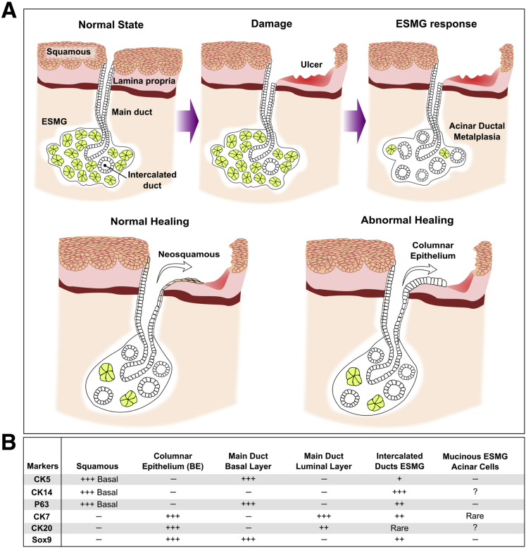
The origin of the progenitor cell for Barrett's esophagus remains a major unsolved mystery. Understanding the source of this progenitor may improve strategies to prevent the development of esophageal adenocarcinoma. Esophageal submucosal glands (ESMGs) and ducts may serve as a potential source of progenitor cells that respond to esophageal injury. Through the use of human histologic and molecular analysis, ESMGs and ducts have been described in physical continuity with areas of columnar esophagus, and shared mutations have been described between ESMG ducts and Barrett's esophagus. Acinar ductal metaplasia, associated with carcinogenesis in other organs, occurs within ESMGs with human esophageal injury and esophageal adenocarcinoma. By using atypical animal models, a squamous epithelial defect well above the gastroesophageal junction healed to columnar epithelium and continuity of ESMG ducts was noted in the new epithelium. Increased proliferation in ESMGs and ducts in response to injury also has been noted in human beings and animals.
Figures
Similar articles
-
Porcine Esophageal Submucosal Gland Culture Model Shows Capacity for Proliferation and Differentiation.Cell Mol Gastroenterol Hepatol. 2017 Aug 4;4(3):385-404. doi: 10.1016/j.jcmgh.2017.07.005. eCollection 2017 Nov. Cell Mol Gastroenterol Hepatol. 2017. PMID: 28936470 Free PMC article.
-
Ductular and proliferative response of esophageal submucosal glands in a porcine model of esophageal injury and repair.Am J Physiol Gastrointest Liver Physiol. 2017 Sep 1;313(3):G180-G191. doi: 10.1152/ajpgi.00036.2017. Epub 2017 Jun 1. Am J Physiol Gastrointest Liver Physiol. 2017. PMID: 28572084 Free PMC article.
-
Transcommitment: Paving the Way to Barrett's Metaplasia.Adv Exp Med Biol. 2016;908:183-212. doi: 10.1007/978-3-319-41388-4_10. Adv Exp Med Biol. 2016. PMID: 27573773 Review.
-
Ductal metaplasia in oesophageal submucosal glands is associated with inflammation and oesophageal adenocarcinoma.Histopathology. 2015 Dec;67(6):771-82. doi: 10.1111/his.12707. Epub 2015 Jun 4. Histopathology. 2015. PMID: 25847432 Free PMC article.
-
Acid, bile, and CDX: the ABCs of making Barrett's metaplasia.Am J Physiol Gastrointest Liver Physiol. 2008 Aug;295(2):G211-8. doi: 10.1152/ajpgi.90250.2008. Epub 2008 Jun 12. Am J Physiol Gastrointest Liver Physiol. 2008. PMID: 18556417 Review.
Cited by
-
Glyco-conjugated bile acids drive the initial metaplastic gland formation from multi-layered glands through crypt-fission in a murine model.PLoS One. 2019 Jul 26;14(7):e0220050. doi: 10.1371/journal.pone.0220050. eCollection 2019. PLoS One. 2019. PMID: 31348796 Free PMC article.
-
Mechanisms and pathophysiology of Barrett oesophagus.Nat Rev Gastroenterol Hepatol. 2022 Sep;19(9):605-620. doi: 10.1038/s41575-022-00622-w. Epub 2022 Jun 7. Nat Rev Gastroenterol Hepatol. 2022. PMID: 35672395 Review.
-
Genetic Mouse Models and Induced Pluripotent Stem Cells for Studying Tracheal-Esophageal Separation and Esophageal Development.Stem Cells Dev. 2020 Aug 1;29(15):953-966. doi: 10.1089/scd.2020.0075. Epub 2020 Jul 2. Stem Cells Dev. 2020. PMID: 32515280 Free PMC article.
-
GATA4 blocks squamous epithelial cell gene expression in human esophageal squamous cells.Sci Rep. 2021 Feb 5;11(1):3206. doi: 10.1038/s41598-021-82557-x. Sci Rep. 2021. PMID: 33547361 Free PMC article.
-
Epithelial cell plasticity: breaking boundaries and changing landscapes.EMBO Rep. 2021 Jul 5;22(7):e51921. doi: 10.15252/embr.202051921. Epub 2021 Jun 6. EMBO Rep. 2021. PMID: 34096150 Free PMC article. Review.
References
-
- van Nieuwenhove Y., Destordeur H., Willems G. Spatial distribution and cell kinetics of the glands in the human esophageal mucosa. Eur J Morphol. 2001;39:163–168. - PubMed
-
- Coad R.A., Woodman A.C., Warner P.J. On the histogenesis of Barrett's oesophagus and its associated squamous islands: a three-dimensional study of their morphological relationship with native oesophageal gland ducts. J Pathol. 2005;206:388–394. - PubMed
-
- Lorinc E., Oberg S. Submucosal glands in the columnar-lined oesophagus: evidence of an association with metaplasia and neosquamous epithelium. Histopathology. 2012;61:53–58. - PubMed
-
- Gillen P., Keeling P., Byrne P.J. Experimental columnar metaplasia in the canine oesophagus. Br J Surg. 1988;75:113–115. - PubMed
Grants and funding
LinkOut - more resources
Full Text Sources
Other Literature Sources
