. 2017 Jul;116(7):1963-1971.
doi: 10.1007/s00436-017-5479-x. Epub 2017 Jun 7.

Origin, differentiation and functional ultrastructure of egg envelopes in the cestode Echinococcus multilocularis Leuckart, 1863 (Cyclophyllidea: Taeniidae)

Affiliations

Origin, differentiation and functional ultrastructure of egg envelopes in the cestode Echinococcus multilocularis Leuckart, 1863 (Cyclophyllidea: Taeniidae)

Zdzisław Świderski et al. Parasitol Res. 2017 Jul.

Abstract

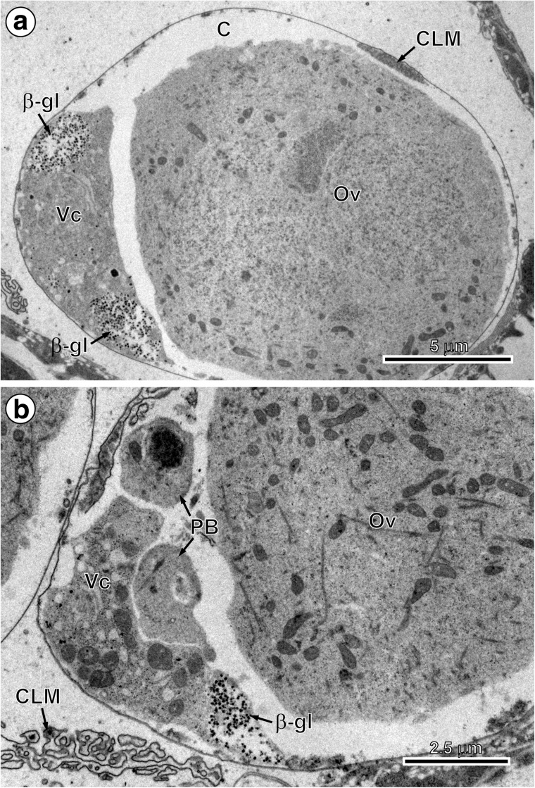
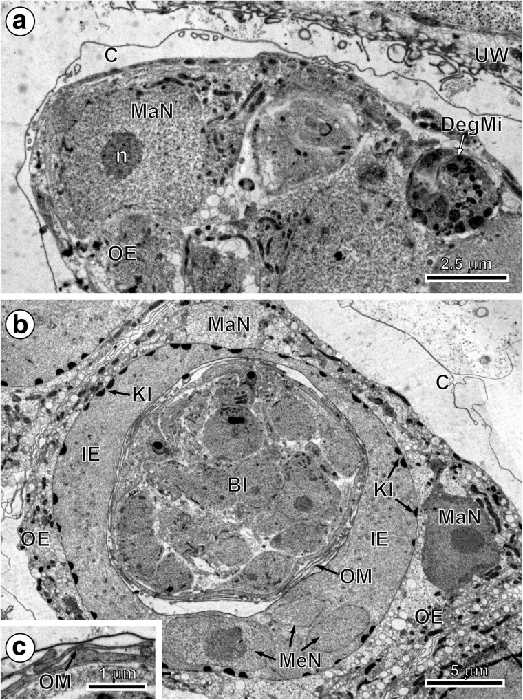
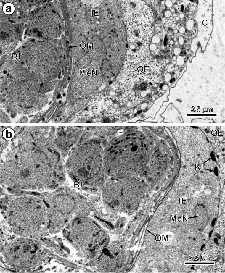
The origin, differentiation and functional ultrastructure of oncospheral or egg envelopes in Echinococcus multilocularis Leuckart, 1863 were studied by transmission electron microscopy (TEM) and cytochemistry. The purpose of our study is to describe the formation of the four primary embryonic envelopes, namely vitelline capsule, outer envelope, inner envelope and oncospheral membrane, and their transformation into the oncospheral or egg envelopes surrounding the mature hexacanth. This transformation takes place in the preoncospheral phase of embryonic development. The vitelline capsule and oncospheral membrane are thin membranes, while the outer and inner envelopes are thick cytoplasmic layers formed by two specific types of blastomeres: the outer envelope by cytoplasmic fusion of two macromeres and the inner envelope by cytoplasmic fusion of three mesomeres. Both outer and inner envelopes are therefore cellular in origin and syncytial in nature. During the advanced phase of embryonic development, the outer and inner envelopes undergo great modifications. The outer envelope remains as a metabolically active layer involved in the storage of glycogen and lipids for the final stages of egg development and survival. The inner envelope is the most important protective layer because of its thick layer of embryophoric blocks that assures oncospheral protection and survival. This embryophore is the principal layer of mature eggs, affording physical and physiological protection for the differentiated embryo or oncosphere, since the outer envelope is stripped from the egg before it is liberated. The embryophore is very thick and impermeable, consisting of polygonal blocks of an inert keratin-like protein held together by a cementing substance. The embryophore therefore assures extreme resistance of eggs, enabling them to withstand a wide range of environmental temperatures and physicochemical conditions.

Keywords: Cestoda; Echinococcus multilocularis; Egg envelopes; Envelopes origin and differentiation; Functional ultrastructure; Taeniidae.


Figures

Fig. 1
Fig. 1
The early stages of embryonic development in Echinococcus multilocularis. a Fertilised oocyte or ovum and adjacent vitellocyte both surrounded by a newly formed vitelline capsule. Note capsule labyrinth membrane complex on the upper surface of the newly formed vitelline capsule and numerous β-glycogen granules in the cytoplasm of vitellocyte. b Part of an early embryo composed of oocyte, remnants of the vitelline cell with β-glycogen particles and two polar bodies. Note infolded labyrinth of capsular membrane in the left lower corner of the micrograph. β-gl β-glycogen, C vitelline capsule, CLM capsule labyrinth membrane, Ov oocyte, PB polar body, Vc vitellocyte
Fig. 2
Fig. 2
Initial stages of the outer and inner envelope formation. a Part of an early embryo adjacent to uterine wall. Note (1) already formed membraneous vitelline capsule surrounding an early embryo composed of several blastomeres of different sizes and (2) a large macromere, situated under the vitelline capsule at the periphery of other blastomeres, which contains predominant nucleus with spherical electron-dense nucleolus, which takes part in the outer envelope formation. b Preoncospheral phase in more advanced stage of embryonic development. Four primary embryonic envelopes (vitelline capsule, outer envelope, inner envelope and oncospheral membrane) are clearly visible. In the outer envelope, note large nucleus of macromeres which predominant nucleolus, numerous elongated mitochondria and higher concentration of free ribosomes. In the inner envelope, note three nuclei of mesomeres, the fusion of which forms the cytoplasm of the syncytial layer. c Detail of the oncospheral membrane. Bl blastomere, C vitelline capsule, DegMi degenerating micromere, IE inner envelope, KI keratin-like protein islands, MaN macromere nucleus, MeN mesomere nucleus, n nucleolus, OE outer envelope, OM oncospheral membrane, UW uterine wall
Fig. 3
Fig. 3
Ultrastructure of embryos in advanced preoncospheral phase of development. a Part of an embryo surrounded by four embryonic envelopes, namely vitelline capsule, outer envelope, inner envelope with nucleus of mesomere and oncospheral membrane. b Part of an embryo composed of numerous blastomeres. Note the initial stage of embryophoric block formation composed of small electron-dense islands of keratin-like protein situated under the outer membrane of the inner envelope which shows the nucleus of mesomere participating in its formation. Bl blastomere, C vitelline capsule, IE inner envelope, KI keratin-like protein islands, MeN mesomere nucleus, OE outer envelope, OM oncospheral membrane
Fig. 4
Fig. 4
Ultrastructure of Echinococcus multilocularis eggs in the middle and more advanced preoncospheral phase of the embryonic development. a Median cross section through the entire egg in the middle phase of preoncospheral development. Note the (1) presence of four embryonic envelopes (vitelline capsule, outer envelope, inner envelope and oncospheral membrane), undergoing differentiation into oncospheral or egg envelopes, (2) presence of a large nucleus of macromere in the outer envelope, (3) presence of a discontinuous layer of flat embryophoric islands of keratin-like protein adjacent to the outer membrane of the inner envelope and (4) evident detachment of the oncospheral membrane from the inner surface of the inner envelope which participated in its formation. b Part of the embryo showing a thin layer of embryophoric blocks and two large flattened mesomere nuclei with prominent electron-dense nucleoli situated in the deeper granular sub-layer of the inner envelope. Bl blastomere, C vitelline capsule, EmB embryophoric blocks of keratin-like protein, IE inner envelope, KI keratin-like protein islands, MaN macromere nucleus, MeN mesomere nucleus, n nucleolus, OE outer envelope, OM oncospheral membrane, UW uterine wall
Fig. 5
Fig. 5
Three external layers of egg envelopes after Thiéry cytochemical test for glycogen. Note (1) several β-glycogen particles and numerous unsaturated lipid droplets in the cytoplasm of the outer envelope, (2) very thick layer of embryophoric blocks in the outer part of the inner envelope with numerous vacuoles and the degenerating small mitochondria embedded between the embryophoric blocks and (3) part of the granular layer of the inner envelope situated under the embryophoric blocks. β-gl β-glycogen, C vitelline capsule, EmB embryophoric blocks of keratin-like protein, GL granular layer of embryophore, L saturated, osmiophobic lipid droplets, OE outer envelope
Fig. 6
Fig. 6
Schematic diagram recapitulating our data on egg envelope ultrastructure with notes on the origin of different layers. C vitelline capsule, EmB embryophoric blocks of keratin-like protein, GL granular layer of embryophore, H oncospheral hooks, HRM hook region membrane, IE inner envelope, MaN macromere nucleus, MeN mesomere nucleus, OE outer envelope, OM oncospheral membrane, OT oncospheral tegument

Similar articles

Cited by

References

    1. Burt MDB. Early morphogenesis in the platyhelminthes with special reference to egg development and development of cestode larvae. Int J Parasitol. 1987;17:241–253. doi: 10.1016/0020-7519(87)90047-6. - DOI - PubMed
    1. Chew MWK. Taenia crassiceps: ultrastructural observations on the oncosphere and associated structures. J Helminthol. 1983;57:101–113. doi: 10.1017/S0022149X00009354. - DOI - PubMed
    1. Chomicz L, Świderski Z. Functional ultrastructure of the oncospheral envelopes of twelve species of the family Hymenolepididae, Ariola, 1899, with aquatic life cycles. Acta Parasitol. 2004;49:177–181.
    1. Chomicz L, Świderski Z, Grytner-Ziêcina B. Ultrastructure of the oncospheral envelopes and their differentiation in the cestode, Dicratotaenia coronula (Dujardin, 1845) (Cyclophyllidea, Hymenolepididae) Acta Parasitol. 1995;40:31–36.
    1. Conn DB. Fine structure of the embryonic envelopes of Oochoristica anolis (Cestoda: Linstowiidae) Z Parasitenkd. 1985;71:639–648. doi: 10.1007/BF00925597. - DOI