. 2017 Jun 8:8:15786.
doi: 10.1038/ncomms15786.

Insights into Hunter syndrome from the structure of iduronate-2-sulfatase

Affiliations

Insights into Hunter syndrome from the structure of iduronate-2-sulfatase

Mykhaylo Demydchuk et al. Nat Commun. .

Abstract

Hunter syndrome is a rare but devastating childhood disease caused by mutations in the IDS gene encoding iduronate-2-sulfatase, a crucial enzyme in the lysosomal degradation pathway of dermatan sulfate and heparan sulfate. These complex glycosaminoglycans have important roles in cell adhesion, growth, proliferation and repair, and their degradation and recycling in the lysosome is essential for cellular maintenance. A variety of disease-causing mutations have been identified throughout the IDS gene. However, understanding the molecular basis of the disease has been impaired by the lack of structural data. Here, we present the crystal structure of human IDS with a covalently bound sulfate ion in the active site. This structure provides essential insight into multiple mechanisms by which pathogenic mutations interfere with enzyme function, and a compelling explanation for severe Hunter syndrome phenotypes. Understanding the structural consequences of disease-associated mutations will facilitate the identification of patients that may benefit from specific tailored therapies.


Conflict of interest statement

The authors declare no competing financial interests.

Figures

Figure 1
Figure 1. X-ray crystal structure of IDS.
(a) Annotated diagram of the IDS sequence, showing the ER signal peptide and propeptide (grey), subdomains (SD) 1 and 2 (light blue and purple, respectively), active-site residues (black), disulfide bridges (yellow) and N-linked glycosylation sites (pink). (b) Natural IDS substrates heparan sulfate (with α1-4 linkage) and dermatan sulfate (α1-3 linkage). The sulfate group removed by IDS is indicated (red). The residue at the +1 position can be either D-glucosamine or D-galactosamine, with variable substituents at C2N, C3, C4 and C6 positions (X=H/SO3; Y=H/SO3/COCH3) (c) Ribbon diagram of IDS coloured by subdomain, shown in three orthogonal views. Disulfide bonds (yellow spheres), calcium ion (grey sphere) and glycans (pink sticks) are shown. The active site is indicated by grey sticks.
Figure 2
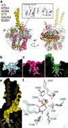
Figure 2. Structural alignment of IDS with other human sulfatase enzymes.
(a) Secondary structure-based superposition of IDS (blue), ARSA (pink), ARSB (green), STS (yellow), GALNS (slate) and SGSH (dark red) in two orthogonal views. Active-site residues are shown as grey sticks. Inset: core structural elements shared by all human sulfatases, showing only conserved regions with backbone Cα RMSD<2.0 Å. (be) Cutaway diagrams of the substrate-binding cleft of IDS, ARSA, ARSB and STS, coloured as above. Active-site residues are shown as grey sticks. (f) Least squares superposition of conserved active-site residues, coloured as above. The metal ions are shown as grey spheres.
Figure 3
Figure 3. Details of IDS active site and substrate-binding cleft.
(a) The substrate-binding cleft is coloured by electrostatic potential at the solvent-accessible surface from red (negative, −4.0 kT e−1) to blue (positive, +4.0 kT e−1). Electrostatic potential was calculated using a pH value of 4.8 when assigning side chain protonation. The catalytic, modified FGS84 residue is shown as sticks. (b) Two orthogonal views of electron density (blue mesh, 2mFODFC, contoured at 0.35 e Å−3) for IDS active-site residues (grey sticks) and the Ca2+ ion (grey sphere). (c) Details of the Ca2+ ion coordination (yellow dashed lines) in the IDS active site. (d) Schematic representation of amino acid side chain interactions with the covalently-modified FGS84 residue. Hydrogen bonds (yellow dashed lines) are shown. (e) Docked structures of 2-sulfo-iduronic acid, two variably sulfated heparan sulfate disaccharides and a dermatan sulfate disaccharide, showing details of the putative substrate-binding region. Hydrogen bonds (dashed yellow lines) are indicated. For solvent-accessible residues situated in the substrate-binding cleft, a graph of average B-factor per residue is shown. The mean B-factor for all IDS non-hydrogen atoms is indicated with a dotted line.
Figure 4
Figure 4. Disease-associated mutations of IDS.
(Top) Annotated diagram of the IDS protein, indicating positions of all known Hunter syndrome missense mutations. All residues that are mutated are shown as sticks (purple) on the structure of IDS, coloured as above. Different mutations have different consequences; for a selection of these a more detailed view is provided (Insets) showing the surrounding region of the structure that is perturbed.

References

    1. Hunter C. A rare disease in two brothers. Proc. R. Soc. Med. 10, 104–116 (1917). - PMC - PubMed
    1. Li P., Bellows A. B. & Thompson J. N. Molecular basis of iduronate-2-sulphatase gene mutations in patients with mucopolysaccharidosis type II (Hunter syndrome). J. Med. Genet. 36, 21–27 (1999). - PMC - PubMed
    1. Lin H. Y. et al.. Incidence of the mucopolysaccharidoses in Taiwan, 1984–2004. Am. J. Med. Genet. A 149A, 960–964 (2009). - PubMed
    1. Baehner F. et al.. Cumulative incidence rates of the mucopolysaccharidoses in Germany. J. Inherit. Metab. Dis. 28, 1011–1017 (2005). - PubMed
    1. Froissart R. et al.. Identification of iduronate sulfatase gene alterations in 70 unrelated Hunter patients. Clin. Genet. 53, 362–368 (1998). - PubMed

Publication types