. 2017 Jun 8:8:15582.
doi: 10.1038/ncomms15582.

Protein-directed ribosomal frameshifting temporally regulates gene expression

Affiliations

Protein-directed ribosomal frameshifting temporally regulates gene expression

Sawsan Napthine et al. Nat Commun. .

Abstract

Programmed -1 ribosomal frameshifting is a mechanism of gene expression, whereby specific signals within messenger RNAs direct a proportion of translating ribosomes to shift -1 nt and continue translating in the new reading frame. Such frameshifting normally occurs at a set ratio and is utilized in the expression of many viral genes and a number of cellular genes. An open question is whether proteins might function as trans-acting switches to turn frameshifting on or off in response to cellular conditions. Here we show that frameshifting in a model RNA virus, encephalomyocarditis virus, is trans-activated by viral protein 2A. As a result, the frameshifting efficiency increases from 0 to 70% (one of the highest known in a mammalian system) over the course of infection, temporally regulating the expression levels of the viral structural and enzymatic proteins.


Conflict of interest statement

The authors declare no competing financial interests.

Figures

Figure 1
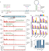
Figure 1. Frameshifting increases dramatically as infection progresses.
(a) Map of the ∼7700-nt EMCV genome. The 5′ and 3′ UTRs are indicated in black and the polyprotein ORF is indicated in pale blue with subdivisions showing mature cleavage products. The overlapping 2B* ORF is indicated in pale pink. (b) Schematic of the ribosome profiling strategy. Each translating ribosome protects ∼30 nt of mRNA. Cells are lysed, treated with RNase I to degrade unprotected mRNA, ribosomes are harvested and the ribosome protected fragments (RPFs) extracted and subjected to high-throughput sequencing. (c) Mutations introduced to prevent PRF. (d) Ribo-Seq RPF densities in reads per million mapped reads (RPM) on WT and SS virus genomes at 4 and 8 h p.i. (e) Ratio of downstream to upstream RPF densities in WT virus divided by the corresponding ratio in SS mutant virus. (f) Phasing of RPFs mapping upstream of 2B*, within the 2B/2B* overlap region, and downstream of 2B*.
Figure 2
Figure 2. An RNA stem–loop is required for frameshifting.
(a) Mutations introduced to prevent PRF (SS) or StopGo-mediated co-translational separation at the C-terminus of 2A (LV) or to destabilize the predicted RNA stem–loop (SL). (b) Ribo-Seq RPF densities in reads per million mapped reads (RPM) on WT and mutant virus genomes at 8 h p.i. WT and SS samples are biological repeats of the 8 h p.i. samples in Fig. 1. (c) Ratio of SS and SS-SL RPF distributions after smoothing with a 31-nt running-mean filter. (d) Phasing of RPFs mapping upstream of 2B*, within the 2B/2B* overlap region, and downstream of 2B*. (e) Ratio of downstream to upstream RPF densities in the different viruses divided by the corresponding ratio in SS mutant virus.
Figure 3
Figure 3. Viral protein 2A binds the stem–loop.
(a) EMSA analysis of binding of 2A to a 64-nt 32P-labelled RNA containing the EMCV PRF signal. Numbers below lanes show fold molar excess of 2A with respect to RNA (10 nM). In lanes BSB, DB and H2O, RNA was incubated alone with band-shift buffer, protein dilution buffer or water, respectively. (b) Phosphorimager quantification of RNA in RNA:protein complexes for (a) and two further repeats. (c) Mutations introduced into the stem–loop for competition assays. (d,e) Unlabelled WT or C46U, G52U or SL3′ mutant competitor RNA was incubated with WT 32P-labelled RNA and 2A (1.8 μM), and analysed by EMSA. Numbers below lanes show fold molar excess of competitor RNA with respect to 32P-labelled WT RNA (10 nM). (f) WT 32P-labelled RNA was incubated with increasing amounts of 2A-mut, and analysed by EMSA. WT 2A was used as a control.
Figure 4
Figure 4. Mutating 2A knocks out frameshifting.
(a) Plaque morphology of WT, SS and 2A-mut viruses on BHK-21 cells (see also ref. 25). (b) Metabolic labelling of BHK-21 cells mock-infected or infected with WT, SS or 2A-mut viruses. Positions of EMCV proteins are indicated. (c) Mean ratio of measureable polyprotein products encoded downstream of the frameshift site to products encoded upstream of the frameshift site, normalized by the SS mutant. Bars show means±s.d. of three biological replicates (green crosses). (d) Time course of 2A expression during WT virus infection assessed by immunoblotting using antibodies to 2A (green) and tubulin (magenta; loading control).
Figure 5
Figure 5. Analysis of the EMCV frameshift signal.
(a) Mutations introduced into the EMCV PRF signal in reporter plasmid pDluc. (be) RNAs derived from FspI-cut plasmid were translated in WG extract in the presence of (b) increasing concentrations of 2A or (d,e) 3 μM 2A or 2A-mut, or 2A dialysis buffer (DB). Products generated by ribosomes that do not frameshift (stop) or that enter the −1 reading frame (−1 FS) are indicated. M and IFC indicate markers and the in-frame control, respectively. Panel c shows the PRF efficiencies for b; the dashed line indicates the 2A concentration used in (d,e).
Figure 6
Figure 6. Ribosomal pausing at the EMCV frameshift signal.
(a) Mutations introduced into the EMCV PRF signal in reporter plasmid pPS0. (be) RNAs derived from AvaII-cut plasmids were translated in WG extract, after 5 min further initiation was halted by the addition of edeine, and aliquots were removed at various times and analysed by SDS–PAGE. Lanes M and C show markers and the expected size of the ribosomal pause product, respectively. Translations were supplemented with 1 μM 2A (bd) or 2A-mut (e). As well as the full-length product and the transient pausing product, a frameshift product is produced for WT RNA only (c).
Figure 7
Figure 7. Comparison of −1 frameshifting stimulators.
(a) Nearly all known cases of −1 PRF are stimulated by an mRNA structure, such as a pseudoknot or stem–loop, separated from the slippery heptanucleotide shift site by a spacer sequence of 5–9 nt, leading to a constant ratio of frameshift to non-frameshift product. (b) In contrast, frameshifting in EMCV is stimulated by a protein:RNA complex positioned at the leading edge of the ribosome when the decoding centre is on the shift site. Increasing levels of viral protein 2A result in a 0 to 70% switch in frameshifting efficiency between early and late timepoints of infection.

Similar articles

Cited by

References

    1. Dinman J. D. Mechanisms and implications of programmed translational frameshifting. Wiley Interdiscip. Rev. RNA 3, 661–673 (2012). - PMC - PubMed
    1. Caliskan N., Peske F. & Rodnina M. V. Changed in translation: mRNA recoding by -1 programmed ribosomal frameshifting. Trends Biochem. Sci. 40, 265–274 (2015). - PMC - PubMed
    1. Atkins J. F., Loughran G., Bhatt P. R., Firth A. E. & Baranov P. V. Ribosomal frameshifting and transcriptional slippage: from genetic steganography & cryptography to adventitious use. Nucleic Acids Res. 44, 7007–7078 (2016). - PMC - PubMed
    1. Belew A. T. et al.. Ribosomal frameshifting in the CCR5 mRNA is regulated by miRNAs and the NMD pathway. Nature 512, 265–269 (2014). - PMC - PubMed
    1. Brierley I., Jenner A. J. & Inglis S. C. Mutational analysis of the "slippery-sequence" component of a coronavirus ribosomal frameshifting signal. J. Mol. Biol. 227, 463–479 (1992). - PMC - PubMed

Publication types

MeSH terms