Review
. 2017 Jun 8:3:17032.
doi: 10.1038/nrdp.2017.32.

Severe congenital neutropenias

Affiliations
Review

Severe congenital neutropenias

Julia Skokowa et al. Nat Rev Dis Primers. .

Abstract

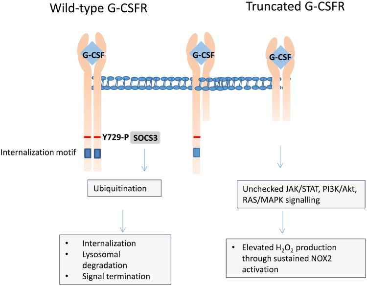
Severe congenital neutropenias are a heterogeneous group of rare haematological diseases characterized by impaired maturation of neutrophil granulocytes. Patients with severe congenital neutropenia are prone to recurrent, often life-threatening infections beginning in their first months of life. The most frequent pathogenic defects are autosomal dominant mutations in ELANE, which encodes neutrophil elastase, and autosomal recessive mutations in HAX1, whose product contributes to the activation of the granulocyte colony-stimulating factor (G-CSF) signalling pathway. The pathophysiological mechanisms of these conditions are the object of extensive research and are not fully understood. Furthermore, severe congenital neutropenias may predispose to myelodysplastic syndromes or acute myeloid leukaemia. Molecular events in the malignant progression include acquired mutations in CSF3R (encoding G-CSF receptor) and subsequently in other leukaemia-associated genes (such as RUNX1) in a majority of patients. Diagnosis is based on clinical manifestations, blood neutrophil count, bone marrow examination and genetic and immunological analyses. Daily subcutaneous G-CSF administration is the treatment of choice and leads to a substantial increase in blood neutrophil count, reduction of infections and drastic improvement of quality of life. Haematopoietic stem cell transplantation is the alternative treatment. Regular clinical assessments (including yearly bone marrow examinations) to monitor treatment course and detect chromosomal abnormalities (for example, monosomy 7 and trisomy 21) as well as somatic pre-leukaemic mutations are recommended.


Figures

Figure 1
Figure 1. Maturation arrest of granulopoiesis in patients with severe congenital neutropenia
A | Normal differentiation of myeloid progenitor cells into neutrophils in the bone marrow and peripheral blood. Right: In a May Grunewald Giemsa stained bone marrow smear of a healthy individual promyelocyte (black arrow) and neutrophil granulocyte (white arrow) are indicated. B | In patients with severe congenital neutropenia, the maturation of granulocytic precursor cells arrests at the stage of promyelocytes and myelocytes, which accumulate in the bone marrow. Right: In a hematoxylin-eosin stained bone marrow smear of a patient with severe congenital neutropenia there are no mature granulocytes and elevated numbers of promyelocytes (arrow). The morphology of promyelocytes is also different: in patients with severe congenital neutropenia, promyelocytes have multiple vacuoles (white arrow), a bulked nucleus and are much bigger than promyelocytes of healthy individuals. ST-HSC, short-term haematopoietic stem cell; MPP, multipotent progenitor; LMPP, lymphoid-primed multipotent progenitor; GMP, granulocyte-macrophage progenitor.
Figure 2
Figure 2. Milestones of the history of severe congenital neutropenia
Severe congenital neutropenia was first described in 1922 (Ref), but referred to as ”agranulocytosis“ or Schultz syndrome ,,. In 1956, Rolf Kostmann described for the first time the autosomal recessive inheritance of familial agranulocytosis and named it as ”infantile genetic agranulocytosis“ Subsequently, the term ”Kostmann syndrome” was used for many years for patients with severe congenital neutropenia. In 1959, a family with dominant inheritance of neutropenia was reported for the first time and in 1970 severe congenital neutropenia was first recognized as a preleukemic syndrome. Recombinant human granulocyte-colony stimulating factor (G-CSF) (for example filgrastim and lenograstim became available for clinical research in 1987 and was approved by the FDA for the treatment of congenital neutropenia in 1993 (Ref). First description of the: GFI1 gene mutation the CXCR4 TCIRG1. First description of the acquired CSF3R mutations in severe congenital neutropenia patients
Figure 3
Figure 3. Genes with germline mutations associated with severe congenital neutropenia
Data based from 650 patients with severe congenital neutropenia registered in the European and North-American Branches of the Severe Chronic Neutropenia International Registry. *Mutations in JAGN1, LAMTOR2, GFI1, LYST, USB1 or mitochondrial DNA.
Figure 4
Figure 4. Main cellular localization of proteins mutated in patients with congenital neutropenia
Mutant proteins are distributed throughout different cellular compartments, including azurophil granules (neutrophil elastase (NE)), plasma membrane (granulocyte-colony stimulating factor receptor (G-CSFR) and C-X-C chemokine receptor type 4 (CXCR4)), endosomes and lysosomes (AP-3 complex subunit beta-1 (AP3B1), lysosomal-trafficking regulator (LYST), Ras-related protein Rab-27A (RAB27A) and ragulator complex protein LAMTOR2 (p14)), mitochondria (HCLS1-associated protein X-1 (HAX-1), tafazzin (TAZ), adenylate kinase 2 (AK2)), endoplasmic reticulum (glucose-6-phosphate exchanger SLC37A4 (G6PT1), glucose-6-phosphatase 3 (G6PC3), protein jagunal homolog 1 (JAGN1) and vacuolar protein sorting-associated protein 13B (VPS13B)), ribosomes (ribosome maturation protein SBDS (SBDS)), nucleus (zinc finger protein Gfi-1 (GFI1) and endothelial transcription factor GATA-2 (GATA2)) and cytoskeleton (Wiskott-Aldrich syndrome protein (WAS) and HCLS1-associated protein X-1 (HAX-1)). The functions of these proteins are described in Table 2.
Figure 5
Figure 5. G-CSFR downstream signalling pathways
Main intracellular pathways involving key signalling molecules activated upon binding of granulocyte-colony stimulating factor (G-CSF) to G-CSF receptor (G-CSFR). Tyrosine-protein kinase JAK2 (JAK-2), which is hyperactivated in myeloid cells of patients with severe congenital neutropenia, leads to phosphorylation and activation of the signal transducer and activator of transcription (STAT) 3 and STAT5A, promoting proliferation of haematopoietic stem cells over granulocytic differentiation; sustained activation of STAT5A has been shown in patients with severe congenital neutropenia. Tyrosine-protein phosphatase non-receptor type 11 (SHP-2) is another component of G-CSF signal transduction; SHP-2 induces dephosphorylation and, thereby, activation of tyrosine-protein kinase Lyn, which in turn in association with another tyrosine kinase Syk phosphorylates and activates haematopoietic lineage cell-specific protein (HCLS1), inducing myeloid differentiation of hematopoietic cells ,,,. Substantially increased levels of SHP-2 have been observed in neutrophils from patients with severe congenital neutropenia. In addition, lymphoid enhancer-binding factor 1 (LEF-1) a transcription factor member of the Wnt signalling pathway, is severely diminished in myeloid progenitors of patients with severe congenital neutropenia. LEF-1 activates the granulocyte-specific transcription factor CCAAT/enhancer binding protein alpha (C/EBPα) and its target genes cyclin D1 and c-MYC, as well as anti-apoptotic factor baculoviral IAP repeat-containing protein 5 (survivin). Expression of all these proteins is severely diminished in myeloid cells of patients with severe congenital neutropenia, along with reduced activity of phosphatidylinositol 3-kinases (PI3K), serine/threonine-protein kinases (Akt), HCLS1-associated protein X-1 (HAX-1) and haematopoietic lineage cell-specific protein (HCLS1),. As a consequence G-CSF therapy activates NAMPT and by this the compensatory emergency granulopoiesis pathway: NAMPT converts nicotinamide (NA) into nicotinamide adenine dinucleotide (NAD+), activating NAD+-dependent protein deacetylases, sirtuins (SIRTs), which in turn activate C/EBPβ. Upregulated pathways are shown in red, downregulated in blue.
Figure 6
Figure 6. Dominant action of G-CSFR truncation mutants leads to sustained proliferation and survival signaling
The native mature G-CSFR protein consists of 836 amino acids, whereas the truncated forms of the CSF3R protein usually vary between 715 and 790 amino acids in length. Truncated G-CSFRs lack the ability to undergo endocytosis, owing to the deletion of a di-leucine motif essential for internalization . Furthermore, they are hampered in endosomal trafficking and lysosomal degradation, owing to the loss of (phospho-tyrosine Y729, the binding site of suppressor of cytokine signalling 3 (SOCS3), which mediates ubiquitination of a critical membrane-proximal lysine residue. G-CSFR forms dimers upon activation by G-CSF. Both wild type G-CSFR homodimers and wild type-truncation mutant G-CSFR heterodimers are subject to normal internalization and lysosomal degradation, whereas the homodimeric truncation mutants accumulate at the plasma membrane. Owing to their prolonged half-life and residence time at the plasma membrane, the G-CSFR truncation mutants act dominantly over the wild type G-CSFR, resulting in an elevated G-CSF-induced proliferative response and sustained activation of signal transducer and activator of transcription 5A (STAT5A) and phosphatidylinositol 3-kinases (PI3K)- serine/threonine-protein kinases (Akt) in myeloid progenitors,. Sustained activation of STAT5 appeared essential for the clonal expansion of haematopoietic stem cells and progenitor cells in mice harbouring the CSF3R-d715 truncation mutation, whereas prolonged PI-3K-Akt activation resulted in the increased production of intracellular reactive oxygen species (ROS), particularly hydrogen peroxide (H2O2), in murine myeloid progenitors via activation of the NADPH oxidase complex 2 (Ref). Increased H2O2 levels inactivate oxidation sensitive phosphatases (such as tyrosine-protein phosphatase non-receptor type 1 (PTPN1) and phosphatidylinositol 3,4,5-trisphosphate 3-phosphatase and dual-specificity protein phosphatase PTEN (PTEN)) that might negatively control G-CSF signalling and cause oxidative damage to proteins, lipids and nucleotides.
Figure 7
Figure 7. Model of leukaemogenesis in severe congenital neutropenia
The development of leukaemia is a multistep process characterized by a series of genetic changes that predispone haematopoietic cells with inherited severe congenital neutropenia-associated mutations to malignant transformation. Prolonged exposure of these cells to high dosages of G-CSF might result in the acquisition of CSF3R mutations that generate truncated G-CSFRs, thereby leading to hypersensitivity to G-CSF, defective G-CSFR signal transduction and clonal proliferation. Subsequently, the acquisition of additional leukemia-associated mutations (for example in RUNX1) or chromosomal aberrations, such as trisomy 21 and monosomy 7, ultimately lead to leukaemogenic transformation.
Figure 8
Figure 8. Severe gingivitis and periodontitis in patients with severe congenital neutropenia
Photographs of buccal cavities of two patients with severe congenital neutropenia caused by ELANE mutations show hypertrophic inflamed reddened gingiva and periodontitis. Peridontitis is common in severe congenital neutropenia patients.
Figure 9
Figure 9. Algorithm for the management of patients with severe congenital neutropenia based on response to granulocyte-colony stimulating factor therapy
More than 90% of patients with severe congenital neutropenia respond well to the G-CSF treatment and reach a sustained absolute neutrophil count >1.0 × 10⁹/L. G-CSF, granulocyte-colony stimulating factor; AML, acute myeloid leukaemia; MDS, myelodysplastic syndrome, abnormalities typical of AML or MDS: trisomy 21, monosomy 7, typical BM morphology.

References

    1. Welte K, Zeidler C, Dale DC. Severe congenital neutropenia. Semin Hematol. 2006;43:189–195. - PubMed
    1. Skokowa J, Germeshausen M, Zeidler C, Welte K. Severe congenital neutropenia: inheritance and pathophysiology. Curr Opin Hematol. 2007;14:22–28. - PubMed
    1. Donadieu J, Beaupain B, Mahlaoui N, Bellanne-Chantelot C. Epidemiology of congenital neutropenia. Hematol Oncol Clin North Am. 2013;27:1–17. - PubMed
    1. Carlsson G, et al. Incidence of severe congenital neutropenia in Sweden and risk of evolution to myelodysplastic syndrome/leukaemia. Br J Haematol. 2012;158:363–369. - PubMed
    1. Lebel A, Yacobovich J, Krasnov T, et al. Genetic analysis and clinical picture of severe congenital neutropenia in Israel. Pediatr Blood Cancer. 2015;62:103–108. - PubMed

MeSH terms

Substances

Supplementary concepts