Review
. 2017 Jun 8;18(6):1233.
doi: 10.3390/ijms18061233.

Interactive Roles of DNA Helicases and Translocases with the Single-Stranded DNA Binding Protein RPA in Nucleic Acid Metabolism

Affiliations
Review

Interactive Roles of DNA Helicases and Translocases with the Single-Stranded DNA Binding Protein RPA in Nucleic Acid Metabolism

Sanket Awate et al. Int J Mol Sci. .

Abstract

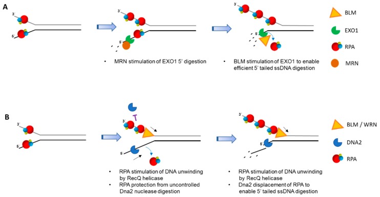
Helicases and translocases use the energy of nucleoside triphosphate binding and hydrolysis to unwind/resolve structured nucleic acids or move along a single-stranded or double-stranded polynucleotide chain, respectively. These molecular motors facilitate a variety of transactions including replication, DNA repair, recombination, and transcription. A key partner of eukaryotic DNA helicases/translocases is the single-stranded DNA binding protein Replication Protein A (RPA). Biochemical, genetic, and cell biological assays have demonstrated that RPA interacts with these human molecular motors physically and functionally, and their association is enriched in cells undergoing replication stress. The roles of DNA helicases/translocases are orchestrated with RPA in pathways of nucleic acid metabolism. RPA stimulates helicase-catalyzed DNA unwinding, enlists translocases to sites of action, and modulates their activities in DNA repair, fork remodeling, checkpoint activation, and telomere maintenance. The dynamic interplay between DNA helicases/translocases and RPA is just beginning to be understood at the molecular and cellular levels, and there is still much to be learned, which may inform potential therapeutic strategies.

Keywords: DNA repair; RPA; Replication Protein A; checkpoint; helicase; replication; telomere; translocase.


Conflict of interest statement

The authors declare no conflict of interest.

Figures

Figure 1
Figure 1
Replication Protein A (RPA) stimulates double-stranded DNA unwinding by the Werner syndrome (WS) helicase-nuclease (WRN) helicase in a manner that is still poorly understood. Two potential mechanisms whereby the physical interaction of RPA with WRN facilitates unwinding of relatively long duplex DNA tracts are depicted. RPA bound to the unwound single-stranded DNA keeps the WRN molecule tethered at the single-stranded/double-stranded DNA junction so that it continues to catalyze unwinding in a processive manner (A). Alternatively, RPA helps to recruit additional WRN helicase molecules in solution to facilitate duplex unwinding (B). The two models are not mutually exclusive and may depend on the DNA structural intermediate or cellular state. Note that positional placement of WRN relative to RPA in (A) and (B) is arbitrary. Adenosine triphosphate (ATP); Adenosine diphosphate (ADP); inorganic phosphate (Pi). RPA heterotrimer is represented by spheres of red (RPA70), blue (RPA32), and yellow (RPA14). Small black arrow indicates directionality of WRN helicase translocation.
Figure 2
Figure 2
RPA regulates SMARCAL1 function at replication forks. RPA binding to single-stranded DNA at replication fork structures with defined polarity modulates ATP-dependent SMARCAL1 DNA translocase in a manner that dictates the fate of stalled (A) or normal (B) replication forks. RPA heterotrimer is represented by spheres of red (RPA70), blue (RPA32), and yellow (RPA14). Physical interaction of RPA32 with SMARCAL1 is not shown. Nascent leading and lagging strands are indicated by green and blue arrows, respectively. See reference [59] and text for details.
Figure 3
Figure 3
RPA enhances multiple ATP-dependent activities catalyzed by FANCJ. RPA stimulates FANCJ unwinding of duplex DNA (A) or resolving G-quadruplex DNA (B). RPA also enhances FANCJ’s ability to bypass a non-translocating strand thymine glycol (C) or displace protein bound to duplex DNA (D). Adenosine triphosphate (ATP); Adenosine diphosphate (ADP); inorganic phosphate (Pi). RPA heterotrimer is represented by spheres of red (RPA70), blue (RPA32), and yellow (RPA14). Small black arrow indicates directionality of FANCJ helicase translocation. Small blue arrow indicates displacement of duplex DNA binding protein. See text for details.
Figure 4
Figure 4
RPA-interacting helicases and DNA nucleases involved in double-strand break (DSB) long range resection generate 3′ single-stranded tails for homologous recombination (HR) repair. After initial trimming by the MRN-CtIPcomplex (not shown), RPA binds single-stranded DNA near ends, thereby preventing inappropriate nucleolytic DNA degradation. (A), MRN, collaborating with RPA, stimulates 5′ to 3′ end-resection by EXO1. Further EXO1 5′ to 3′ end-resection is enhanced by Bloom’s syndrome helicase (BLM) protein interaction with EXO1 that is independent of BLM helicase activity. In the process, RPA becomes unbound from the degraded end. (B), RPA is poised to stimulate DNA unwinding by BLM and/or WRN helicases, creating longer single-stranded DNA. In addition, RPA protects single-stranded DNA with a free 3′ end from being degraded by Dna2 nuclease activity. The 5′ to 3′ helicase-nuclease DNA2 is directed by RPA binding to degrade 5′-ended single-stranded DNA. In both (A,B), the long 3′ single-stranded DNA tails are used for RAD51-mediated strand pairing with complementary DNA strand of recipient duplex for HR repair (not shown). RPA heterotrimer is represented by spheres of red (RPA70), blue (RPA32), and yellow (RPA14). Small black arrow indicates directionality of BLM/WRN helicase translocation.
Figure 5
Figure 5
Small molecule drugs that target helicase-RPA interactions. Biologically active small molecules that bind to a DNA helicase, thereby inhibiting its unwinding function directly (A) or disrupting its physical interaction with RPA (B), may be used to target helicase-dependent pathways of DNA metabolism in human cells. Alternatively, RPA-interacting compounds that interfere with single-stranded DNA binding (C) or physical interaction with a helicase (D) may compromise helicase-dependent pathways. RPA heterotrimer is represented by spheres of red (RPA70), blue (RPA32), and yellow (RPA14). Small black arrow indicates directionality of WRN helicase translocation. Small purple T symbol represents inhibition of designated interaction or action of WRN or RPA. Note that small molecule inhibition mechanisms are not necessarily mutually exclusive.

References

    1. Lohman T.M., Tomko E.J., Wu C.G. Non-hexameric DNA helicases and translocases: Mechanisms and regulation. Nat. Rev. Mol. Cell. Biol. 2008;9:391–401. doi: 10.1038/nrm2394. - DOI - PubMed
    1. Patel S.S., Donmez I. Mechanisms of helicases. J. Biol. Chem. 2006;281:18265–18268. doi: 10.1074/jbc.R600008200. - DOI - PubMed
    1. Singleton M.R., Dillingham M.S., Wigley D.B. Structure and mechanism of helicases and nucleic acid translocases. Annu. Rev. Biochem. 2007;76:23–50. doi: 10.1146/annurev.biochem.76.052305.115300. - DOI - PubMed
    1. Mendoza O., Bourdoncle A., Boule J.B., Brosh R.M., Jr., Mergny J.L. G-quadruplexes and helicases. Nucleic. Acids Res. 2016;44:1989–2006. doi: 10.1093/nar/gkw079. - DOI - PMC - PubMed
    1. Sharma S. Non-B DNA secondary structures and their resolution by RecQ helicases. J. Nucleic Acids. 2011;2011:724215. doi: 10.4061/2011/724215. - DOI - PMC - PubMed

MeSH terms

LinkOut - more resources