. 2017 Jun 8;6(6):58.
doi: 10.3390/jcm6060058.

An Effective, Versatile, and Inexpensive Device for Oxygen Uptake Measurement

Affiliations

An Effective, Versatile, and Inexpensive Device for Oxygen Uptake Measurement

Paule Bénit et al. J Clin Med. .

Abstract

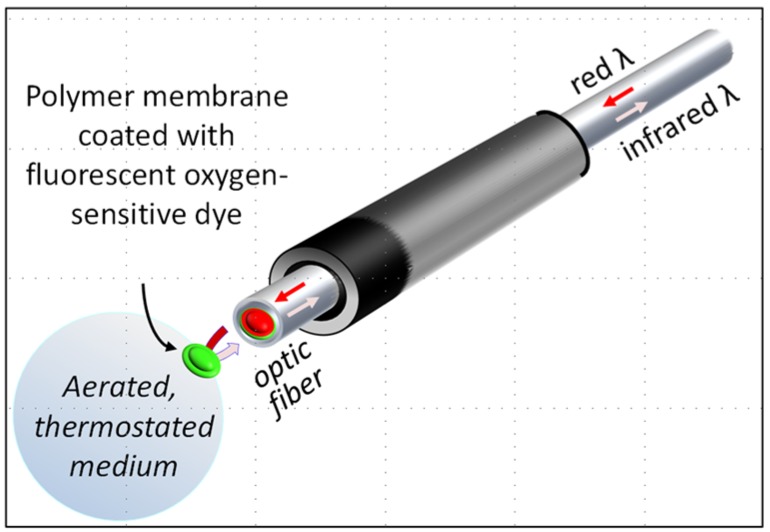
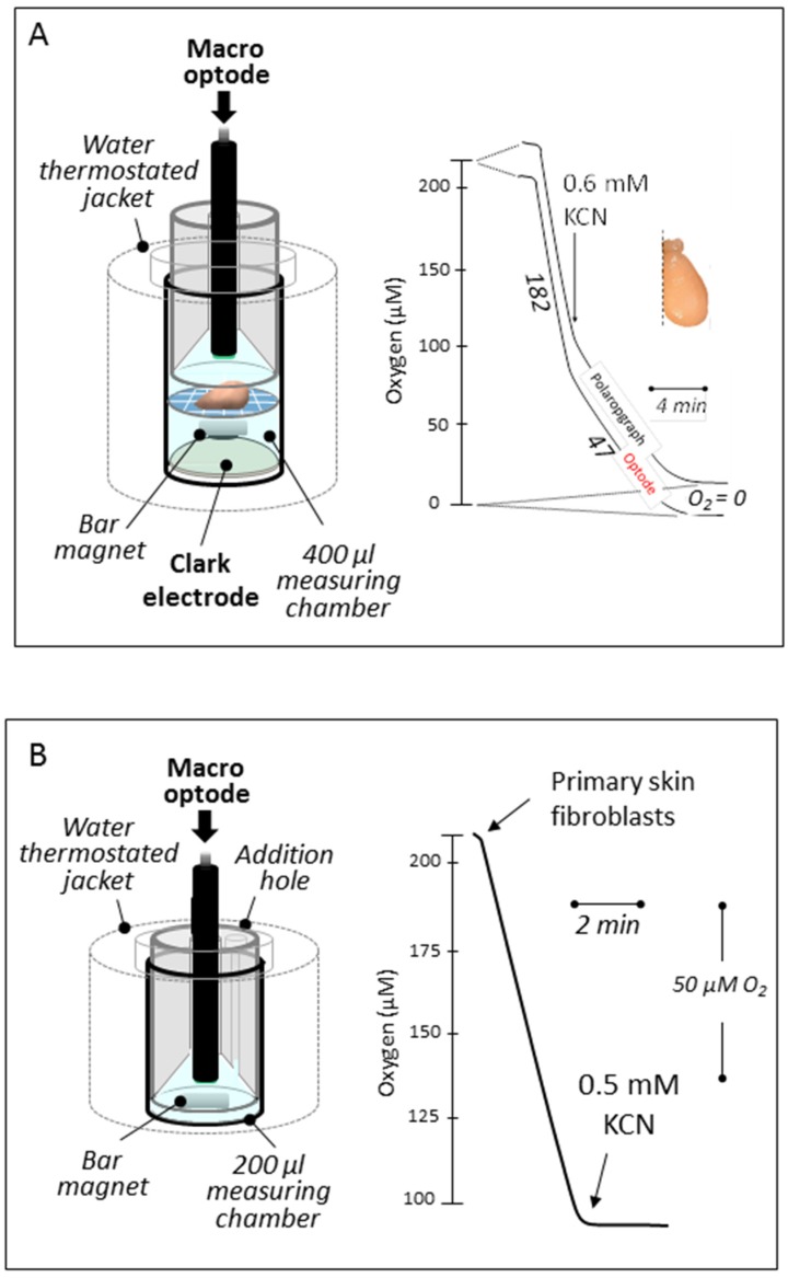
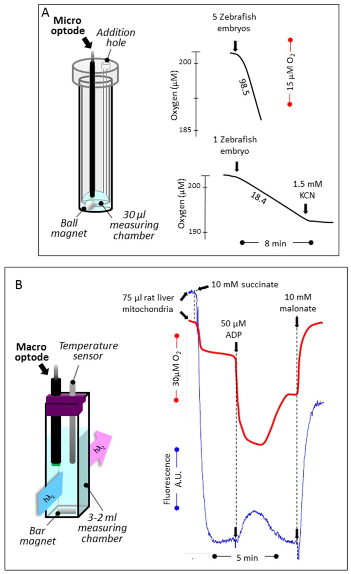
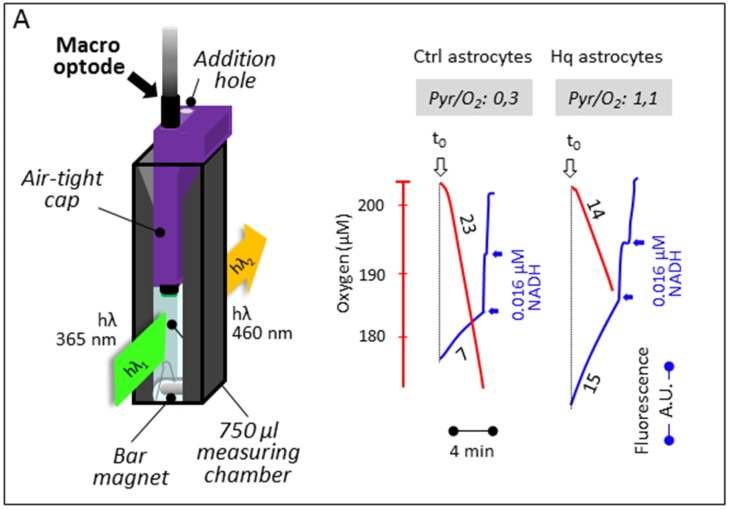
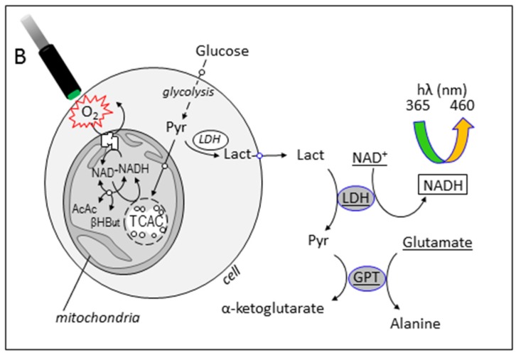
In the last ten years, the use of fluorescent probes developed to measure oxygen has resulted in several marketed devices, some unreasonably expensive and with little flexibility. We have explored the use of the effective, versatile, and inexpensive Redflash technology to determine oxygen uptake by a number of different biological samples using various layouts. This technology relies on the use of an optic fiber equipped at its tip with a membrane coated with a fluorescent dye (www.pyro-science.com). This oxygen-sensitive dye uses red light excitation and lifetime detection in the near infrared. So far, the use of this technology has mostly been used to determine oxygen concentration in open spaces for environmental studies, especially in aquatic media. The oxygen uptake determined by the device can be easily assessed in small volumes of respiration medium and combined with the measurement of additional parameters, such as lactate excretion by intact cells or the membrane potential of purified mitochondria. We conclude that the performance of by this technology should make it a first choice in the context of both fundamental studies and investigations for respiratory chain deficiencies in human samples.

Keywords: glycolysis; mitochondriopathy; oxygen uptake; respiration assay.


Conflict of interest statement

The authors declare no conflict of interest.

Figures

Figure 1
Figure 1
The optode device. The tip of the optic fiber (in red) is covered by a polymer membrane coated with a fluorescent oxygen-sensitive dye (in green) fixed to the optic fiber with silicone glue. The optic fiber receives red light (excitation) from, and re-emits infrared light (emission) to, an analyzer box that can be connected to a personal computer. The fluorescence of the dye is proportional to its oxygen-dependent oxidation state, which is fully reversible. Time-dependent variation of the infrared emission reflects variation of the oxygen at the membrane surface. By inserting the tip of the optic fiber into any aerated medium, it appears possible to determine at any time the oxygen tension in the medium and so to estimate oxygen consumption in the medium.
Figure 2
Figure 2
In vitro oxygen uptake by tissue sample and cells in suspension. (A) Both the Hansatech polarographic device (bottom) and the FireSting optode work simultaneously allowing to record strictly similar rates of oxygen uptake (about 25% resistant to 0.6 mM cyanide) by a mouse brain hemisphere place on a nylon net at mid-height in 400 μL of respiratory medium A (see Material and Methods). (B) Fully cyanide-sensitive human primary fibroblast respiration (about 1 × 10⁶ cells for 50 μM O2/min) recorded in 200 μL of respiratory medium A. Numbers along the traces are nmol/min/mg protein.
Figure 3
Figure 3
Respiration of Zebrafish embryos, and oxygen tension coupled to membrane potential determination by rat liver mitochondria. (A) Cyanide-sensitive respiration of Zebrafish embryos (1 and 5) measured at ambient temperature (20 °C) using a micro-optode in 30 μL of PBS (linear rates observed for 10 min). Notice the spherical magnetic stirrer avoiding to hurt the embryos. (B) Using an open layout, oxygen level changes linked to mitochondrial substrate oxidation was measured concomitantly to the membrane potential. Oxygen was measured using the macro-optode placed in a 3 mL quartz cell thermostated at 38 °C and magnetically stirred. Oxygen uptake (red trace) was started by the addition of succinate followed by the addition of a limiting amount of ADP (decreased level of oxygen, due to high rate of consumption; oxidation state 3 [20]), the exhaustion of which exhaustion in a higher oxygen level (oxidation state 4). The addition of malonate (a long established inhibitor of the succinate dehydrogenase [21]) fully inhibited oxygen uptake, and the level of oxygenation of the medium came back to initial value by re-equilibration with air. The membrane potential measured simultaneously (blue trace) rose upon succinate addition (quenching of rhodamine fluorescence) to drop down upon the ADP addition. After ADP exhaustion, the membrane potential rose again, while adding malonate worked to abolish most of it. Numbers along the traces are nmol/min/mg protein.
Figure 4
Figure 4
Respiration and lactate excretion by mouse-cultured astrocytes. (A) The macro-optode fitted to a magnetically stirred, 37.5 °C-thermostated quartz-cell by a closed cap (yet allowing for the addition of substrates and inhibitors) measures oxygen uptake due to cyanide-sensitive respiration (red traces) by control astrocytes or astrocytes prepared from the CI-defective Harlequin mice. The concomitant fluorometric determination of NADH (blue traces) allows for a determination of the rate of the excretion of lactate, thanks to its conversion to pyruvate brought about by added lactate dehydrogenase in the presence of added NAD+. Numbers along the traces are nmol/min/mg protein. (B) The additional presence of glutamate and glutamate transaminase avoided inhibition of the LDH reaction by accumulated pyruvate.
Figure 4
Figure 4
Respiration and lactate excretion by mouse-cultured astrocytes. (A) The macro-optode fitted to a magnetically stirred, 37.5 °C-thermostated quartz-cell by a closed cap (yet allowing for the addition of substrates and inhibitors) measures oxygen uptake due to cyanide-sensitive respiration (red traces) by control astrocytes or astrocytes prepared from the CI-defective Harlequin mice. The concomitant fluorometric determination of NADH (blue traces) allows for a determination of the rate of the excretion of lactate, thanks to its conversion to pyruvate brought about by added lactate dehydrogenase in the presence of added NAD+. Numbers along the traces are nmol/min/mg protein. (B) The additional presence of glutamate and glutamate transaminase avoided inhibition of the LDH reaction by accumulated pyruvate.

References

    1. Galluzzi L., Kepp O., Trojel-Hansen C., Kroemer G. Mitochondrial control of cellular life, stress, and death. Circ. Res. 2012;111:1198–1207. doi: 10.1161/CIRCRESAHA.112.268946. - DOI - PubMed
    1. Rustin P., Chretien D., Bourgeron T., Gerard B., Rotig A., Saudubray J.M., Munnich A. Biochemical and molecular investigations in respiratory chain deficiencies. Clin. Chim. Acta. 1994;228:35–51. doi: 10.1016/0009-8981(94)90055-8. - DOI - PubMed
    1. Turnbull D.M., Rustin P. Genetic and biochemical intricacy shapes mitochondrial cytopathies. Neurobiol. Dis. 2016;92:55–63. doi: 10.1016/j.nbd.2015.02.003. - DOI - PubMed
    1. Benit P., Letouze E., Rak M., Aubry L., Burnichon N., Favier J., Gimenez-Roqueplo A.P., Rustin P. Unsuspected task for an old team: Succinate, fumarate and other krebs cycle acids in metabolic remodeling. Biochim. Biophys. Acta. 2014;1837:1330–1337. doi: 10.1016/j.bbabio.2014.03.013. - DOI - PubMed
    1. Golpich M., Amini E., Mohamed Z., Azman Ali R., Mohamed Ibrahim N., Ahmadiani A. Mitochondrial dysfunction and biogenesis in neurodegenerative diseases: Pathogenesis and treatment. CNS Neurosci. Ther. 2017;23:5–22. doi: 10.1111/cns.12655. - DOI - PMC - PubMed

LinkOut - more resources