. 2017 Nov;101(11):2731-2738.
doi: 10.1097/TP.0000000000001814.

DHRS9 Is a Stable Marker of Human Regulatory Macrophages

Affiliations

DHRS9 Is a Stable Marker of Human Regulatory Macrophages

Paloma Riquelme et al. Transplantation. 2017 Nov.

Abstract

Background: The human regulatory macrophage (Mreg) has emerged as a promising cell type for use as a cell-based adjunct immunosuppressive therapy in solid organ transplant recipients. In this brief report, dehydrogenase/reductase 9 (DHRS9) is identified as a robust marker of human Mregs.

Methods: The cognate antigen of a mouse monoclonal antibody raised against human Mregs was identified as DHRS9 by immunoprecipitation and MALDI-MS sequencing. Expression of DHRS9 within a panel of monocyte-derived macrophages was investigated by quantitative PCR, immunoblotting and flow cytometry.

Results: DHRS9 expression discriminated human Mregs from a panel of in vitro derived macrophages in other polarisation states. Likewise, DHRS9 expression distinguished Mregs from a variety of human monocyte-derived tolerogenic antigen-presenting cells in current development as cell-based immunotherapies, including Tol-DC, Rapa-DC, DC-10, and PGE2-induced myeloid-derived suppressor cells. A subpopulation of DHRS9-expressing human splenic macrophages was identified by immunohistochemistry. Expression of DHRS9 was acquired gradually during in vitro development of human Mregs from CD14 monocytes and was further enhanced by IFN-γ treatment on day 6 of culture. Stimulating Mregs with 100 ng/mL lipopolysaccharide for 24 hours did not extinguish DHRS9 expression. Dhrs9 was not an informative marker of mouse Mregs.

Conclusion: DHRS9 is a specific and stable marker of human Mregs.


Conflict of interest statement

J.A.H. and E.K.G. are the named inventors on European Patent Office (EPO) filing 16159985.6-1402 dated 11.03.2016, “Immunoregulatory cells and methods for their production.” All other authors declare no conflict-of-interest.

Figures
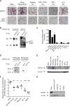
FIGURE 2
FIGURE 2
DHRS9 expression distinguishes human Mregs from monocyte-derived macrophages and DCs. A, In immunocytochemistry, the ASOT1 mAb recognized an antigen expressed by Mregs, but not comparator macrophages. B, An antigen of ~35 kDa was specifically immunoprecipitated by ASOT1 and was subsequently identified by MALDI-MS as DHRS9. C, Strong DHRS9 mRNA expression was detected in Mregs, but not comparator macrophage types (n = 6; mean ± SD). D, ASOT1 precipitated an antigen which was also recognized by an anti-DHRS9 rabbit pAb (generated in-house) and a mouse mAb (clone 3C6, Abnova), confirming that ASOT1 recognizes DHRS9. E, Immunoblotting with our custom-made rabbit anti-DHRS9 pAb demonstrated that DHRS9 protein expression distinguishes Mregs from comparator macrophages. F, DHRS9 mRNA expression distinguished human Mregs from a panel of tolerogenic monocyte-derived therapeutic cell products. G, DHRS9 protein was detected in human Mregs but not other tolerogenic monocyte-derived therapeutic cell products using a commercial rabbit anti-DHRS9 pAb (ab98155, Abcam).
FIGURE 1
FIGURE 1
Comparative phenotyping of Mregs and other human macrophages. Low or absent expression of CD14, CD16, TLR2 and CD163 discriminated human Mregs from a panel of differently stimulated human monocyte-derived macrophages, including resting Mφ, LPS + IFNγ-stimulated Mφ, IL-4-stimulated Mφ, immunoglobulin (Ig)-stimulated Mφ and GC-stimulated Mφ. Values represent mean ± SD of n = 6 donors.
FIGURE 3
FIGURE 3
Factors affecting DHRS9 expression in human macrophages. A, DHRS9 mRNA expression increased steadily over 7 days of culture as human monocytes transitioned to Mregs and was further induced by IFN-γ stimulation on day 6 (n = 6; mean ± SD). B, Pattern of DHRS9 expression in a publicly available microarray data set encompassing 29 differently stimulated human monocyte-derived macrophages (Xue, J. et al, Immunity; GEO accession: GSE47189). More than twofold upregulation of DHRS9 was elicited by treatment with either 1 μM dexamethasone or 200 IU/mL IFN-γ. HDL, high-density lipoprotein; IC, immune complex; P3C, Pam3CSK4; PGE2, Prostaglandin E2; LA, lauric acid; OA, oleic acid; LiA, linoleic acid; SA, steric acid; PA, palmitic acid; TPP, TNF + PGE2 + P3C. C, Optimization of an intracellular staining method for detection of DHRS9 expression in human macrophages using PE-conjugated anti-DHRS9 mAb clone 3C6. D, Quantification of DHRS9 expression in human Mregs, IFN-γ Mφ and resting Mφ by flow cytometry.
FIGURE 4
FIGURE 4
Human Mregs express enzymes involved in retinoid metabolism. A, A schematic overview of retinoid metabolism. DHRS9 is a member of the SDR family of NAD(P)(H)-dependent oxidoreductases that catalyse interconversion of retinol and retinal. B, Mregs expressed ALDH1A1, ALDH1A2 and BCO2 mRNA, as well as the RA-responsive gene, CD1D (n = 3).
FIGURE 5
FIGURE 5
Identification of DHRS9+ Mφ in the subcapsular red pulp of human spleen. Immunohistochemical staining of 3 μm, H&E-counterstained sections of fixed, paraffin-embedded human spleen revealed a population of DHRS9+ macrophages. A, DHRS9+ Mφ detected with mAb 3C6 appeared to be most prevalent in the subcapsular red pulp. Original magnification, 20×. B, Negative control. Original magnification, 20×. C, A high density of DHRS9+ macrophages were detected in the subcapsular splenic red pulp using mAb 3C6. Original magnification, 40×. D, DHRS9 staining with mAb 3C6 was restricted to cytoplasm of cells with typical macrophage morphology. Original magnification, 40×.
FIGURE 6
FIGURE 6
DHRS9 is not a marker of mouse Mregs. Microarray analysis of SDR-family gene expression in monocyte-derived macrophages and DCs from C57BL/6 mice (Riquelme, P. et al, Molecular Therapy; GEO accession: GSE32690). No differential expression of DHRS9 or other SDR family members was observed between mouse Mregs and comparator cell types.

Comment in

References

    1. Hutchinson JA, Geissler EK. Now or never? The case for cell-based immunosuppression in kidney transplantation. Kidney Int. 2015;87:1116–1124. - PubMed
    1. Hutchinson JA, Riquelme P, Geissler EK, et al. Human regulatory macrophages. Methods Mol Biol. 2011;677:181–192. - PubMed
    1. Hutchinson JA, Riquelme P, Sawitzki B, et al. Cutting edge: immunological consequences and trafficking of human regulatory macrophages administered to renal transplant recipients. J Immunol. 2011;187:2072–2078. - PubMed
    1. Ahrens N, Riquelme P, Bach C, et al. Donor-specific anti-HLA antibodies present in pooled human serum do not prevent development of human Mreg_UKR from monocytes in culture. Transplantation. 2017;101:e188–e190. - PubMed
    1. Hutchinson JA. Macrophages in transplantation. Transplantation. 2015;99:898–899. - PubMed

Publication types

MeSH terms