Comparative Study
. 2017 Jun 8;13(6):e1006343.
doi: 10.1371/journal.ppat.1006343. eCollection 2017 Jun.

NS5A inhibitors unmask differences in functional replicase complex half-life between different hepatitis C virus strains

Affiliations
Comparative Study

NS5A inhibitors unmask differences in functional replicase complex half-life between different hepatitis C virus strains

Tiffany Benzine et al. PLoS Pathog. .

Abstract

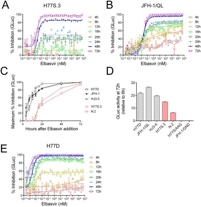
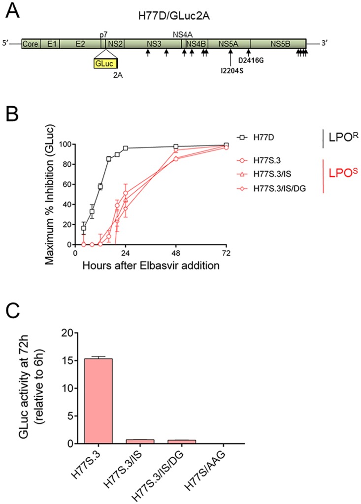
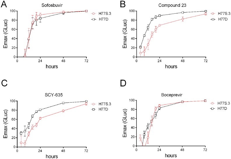
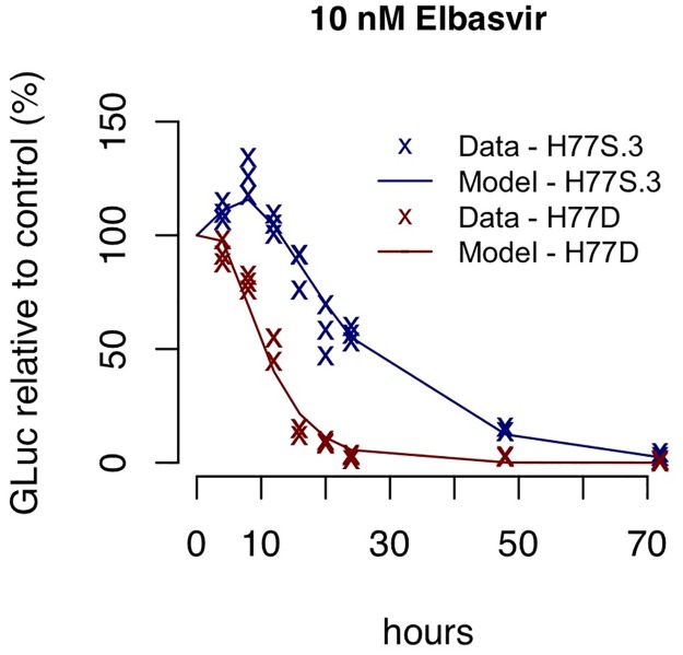
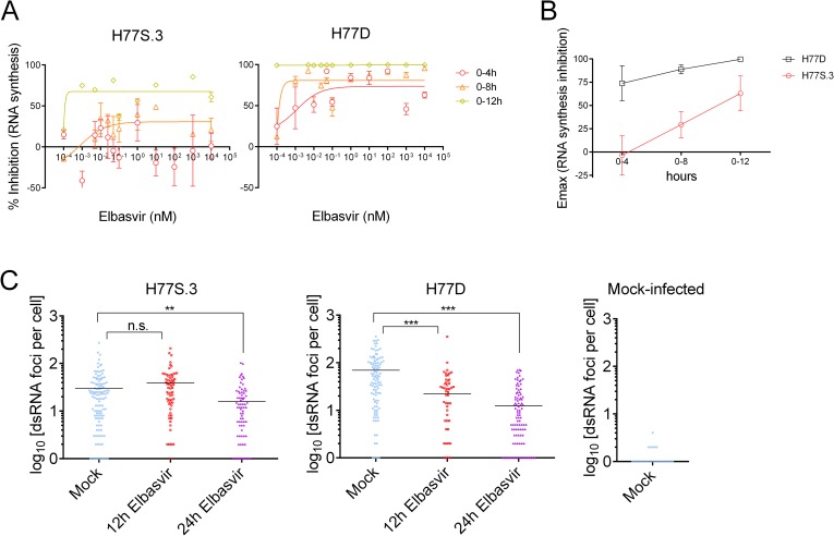
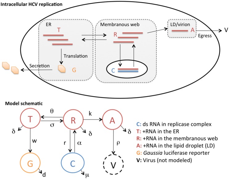
Hepatitis C virus (HCV) RNA is synthesized by the replicase complex (RC), a macromolecular assembly composed of viral non-structural proteins and cellular co-factors. Inhibitors of the HCV NS5A protein block formation of new RCs but do not affect RNA synthesis by pre-formed RCs. Without new RC formation, existing RCs turn over and are eventually lost from the cell. We aimed to use NS5A inhibitors to estimate the half-life of the functional RC of HCV. We compared different cell culture-infectious strains of HCV that may be grouped based on their sensitivity to lipid peroxidation: robustly replicating, lipid peroxidation resistant (LPOR) viruses (e.g. JFH-1 or H77D) and more slowly replicating, lipid peroxidation sensitive (LPOS) viruses (e.g. H77S.3 and N.2). In luciferase assays, LPOS HCV strains declined under NS5A inhibitor therapy with much slower kinetics compared to LPOR HCV strains. This difference in rate of decline was not observed for inhibitors of the NS5B RNA-dependent RNA polymerase suggesting that the difference was not simply a consequence of differences in RNA stability. In further analyses, we compared two isoclonal HCV variants: the LPOS H77S.3 and the LPOR H77D that differ only by 12 amino acids. Differences in rate of decline between H77S.3 and H77D following NS5A inhibitor addition were not due to amino acid sequences in NS5A but rather due to a combination of amino acid differences in the non-structural proteins that make up the HCV RC. Mathematical modeling of intracellular HCV RNA dynamics suggested that differences in RC stability (half-lives of 3.5 and 9.9 hours, for H77D and H77S.3, respectively) are responsible for the different kinetics of antiviral suppression between LPOS and LPOR viruses. In nascent RNA capture assays, the rate of RNA synthesis decline following NS5A inhibitor addition was significantly faster for H77D compared to H77S.3 indicating different half-lives of functional RCs.


Conflict of interest statement

I have read the journal's policy and the authors of this manuscript have the following conflicts of interest: SML has received royalties from AbbVie and grant funding from Merck and Gilead Sciences. DRM has received grant funding from AbbVie and Gilead Sciences. All other authors have no competing interests.

Figures

Fig 1
Fig 1. Kinetics of HCV inhibition in infected cell culture following addition of an NS5A inhibitor, monitored using different GLuc reporter viruses.
(A) Inhibition of the gt1a LPOS virus H77S.3/GLuc2A by elbasvir. (B) Inhibition of the gt2a LPOR virus JFH-1/QL/GLuc2A by elbasvir. (C) Maximum % inhibition (Emax) of GLuc activity at different time points after addition of elbasvir to Huh7.5 cells infected with different cell culture infectious HCV genomes. LPOS viruses H77S.3/GLuc2A (gt1a) and N.2/GLuc2A (gt1b) are plotted in red. LPOR viruses H77D/GLuc2A (gt1a), JFH-1/QL/GLuc2A (gt2a) or HJ3-5/GLuc2A (gt1a/2a chimera). (D) Inhibition of the gt1a LPOR virus H77D-GLuc2A by elbasvir. (E) Fitness of different virus genomes used in (C) at 72 h post electroporation. Data shown represents GLuc activity relative to GLuc activity at 6 hours post electroporation to normalize for transfection efficiency. The replication incompetent reporter viruses H77S/AAG/GLuc2A and JFH-1/GND/GLuc2A contain point mutations in the NS5B polymerase and are included as mock-transfection controls.
Fig 2
Fig 2. Differences in kinetics of virus inhibition in H77S.3- vs H77D–infected cells following addition of an NS5A inhibitor are not due to differences in NS5A sequence.
(A) Diagram of the H77S.3 genome showing positions of the 12 amino acids that differ between H77S.3 and H77D. The two amino acid changes in the NS5A coding region (I2204S and D2416G) are highlighted. (B) Maximum % inhibition (Emax) at different time points after addition of elbasvir to Huh7.5 cells infected with either H77S.3/GLuc2A, H77D/GLuc2A or H77S.3/GLuc2A carrying either the I2204S mutation alone (H77S.3/IS) or in combination with D2416G (H77S.3/IS/DG). (C) Fitness of different virus genomes used in (B) at 72 h post electroporation. Data shown represents GLuc activity relative to GLuc activity at 6 h post electroporation to normalize for transfection efficiency. The replication incompetent reporter virus H77S/AAG/GLuc2A contains point mutations in the NS5B polymerase and serves as a mock-transfection control.
Fig 3
Fig 3. Differences in kinetics of virus inhibition in H77S.3- vs H77D-infected cells are observed with host-targeting antiviral drugs that block membranous web formation but not with the NS5B RdRP inhibitor sofosbuvir or the NS3/4A protease inhibitor boceprevir.
Maximum % inhibition (Emax) at different time points after addition of (A) sofosbuvir, (B) Compound 23: a small molecule inhibitor of PI4KIIIα, (C) SCY-635: a cyclophilin inhibitor, or (D) boceprevir, to Huh7.5 cells infected with H77S.3/GLuc2A or H77D/GLuc2A.
Fig 4
Fig 4. Results of the mathematical model fitted to data measuring the decline of GLuc-reporter activity from the H77S.3 or H77D variants under 10nM elbasvir treatment.
Data and simulation using best fit parameter values are shown as ‘x’s and lines, respectively. The data and simulation shown here represent the 10nM elbasvir treatment from S5 and S8 Figs. Here, they are overlaid on the same graph to highlight the transient increase in GLuc activity observed at 4-8h following NS5A inhibitor addition to cell cultures infected with H77S.3/GLuc2A but not H77D/GLuc2A.
Fig 5
Fig 5. Slower kinetics of RNA synthesis inhibition by an NS5A inhibitor for H77S.3 compared to H77D.
(A) Inhibition of RNA synthesis measured in cultures of Huh7.5 cells infected with either H77S.3 (left panel) or H77D (right panel) following treatment with elbasvir. (B) Maximum % inhibition (Emax) of RNA synthesis at different time points after addition of elbasvir to Huh7.5 cells infected with either H77S.3 or H77D. (C) Measurements of dsRNA foci per cell in Huh7.5 cells infected with either H77S.3 (left panel), H77D (middle panel) or mock-infected cells (right panel). Cells were either mock-treated or treated with 10x EC90 elbasvir for 12 or 24 h before being fixed and stained using a dsRNA specific antibody. Foci of dsRNA per cell were quantified using MetaMorph Image analysis software. Treatment groups were analyzed by Kolmogorov-Smirnoff test (n.s.: not significant, ** p = 0.003, ***p<0.001).
Fig 6
Fig 6. Schematics for intracellular HCV replication dynamics considered in this study and the corresponding mathematical model.
We consider the dynamics of positive strand HCV RNAs (+RNAs) in the ER (T), in the membranous web (R) and in the lipid droplets (A), the replicase complex (C) and the extracellular Gaussia luciferase (GLuc) proteins (G). See text for detailed explanations.

References

    1. Gao M, Nettles RE, Belema M, Snyder LB, Nguyen VN, Fridell RA, et al. Chemical genetics strategy identifies an HCV NS5A inhibitor with a potent clinical effect. Nature. 2010;465(7294):96–100. 10.1038/nature08960 - DOI - PMC - PubMed
    1. McGivern DR, Masaki T, Williford S, Ingravallo P, Feng Z, Lahser F, et al. Kinetic analyses reveal potent and early blockade of hepatitis C virus assembly by NS5A inhibitors. Gastroenterology. 2014;147(2):453–62 e7. 10.1053/j.gastro.2014.04.021 - DOI - PMC - PubMed
    1. Guedj J, Dahari H, Rong L, Sansone ND, Nettles RE, Cotler SJ, et al. Modeling shows that the NS5A inhibitor daclatasvir has two modes of action and yields a shorter estimate of the hepatitis C virus half-life. Proc Natl Acad Sci U S A. 2013;110(10):3991–6. Epub 2013/02/23. 1203110110 [pii] 10.1073/pnas.1203110110 . - DOI - PMC - PubMed
    1. Berger C, Romero-Brey I, Radujkovic D, Terreux R, Zayas M, Paul D, et al. Daclatasvir-like inhibitors of NS5A block early biogenesis of hepatitis C virus-induced membranous replication factories, independent of RNA replication. Gastroenterology. 2014;147(5):1094–105 e25. 10.1053/j.gastro.2014.07.019 - DOI - PubMed
    1. Reiss S, Rebhan I, Backes P, Romero-Brey I, Erfle H, Matula P, et al. Recruitment and activation of a lipid kinase by hepatitis C virus NS5A is essential for integrity of the membranous replication compartment. Cell host & microbe. 2011;9(1):32–45. 10.1016/j.chom.2010.12.002 - DOI - PMC - PubMed

Publication types

MeSH terms

Substances