. 2017 Jun 8;12(6):e0178995.
doi: 10.1371/journal.pone.0178995. eCollection 2017.

Effects of PPARα inhibition in head and neck paraganglioma cells

Affiliations

Effects of PPARα inhibition in head and neck paraganglioma cells

Rosalba Florio et al. PLoS One. .

Abstract

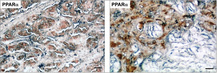
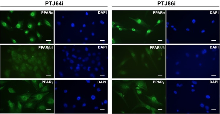
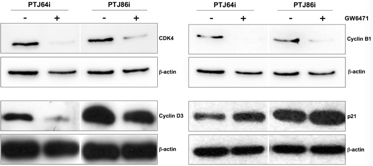
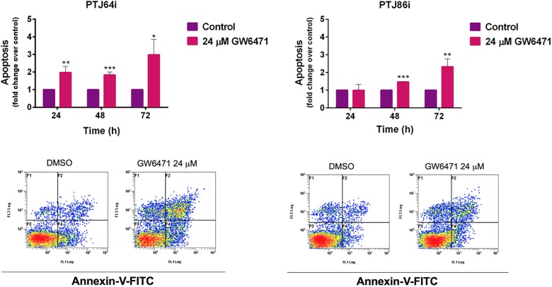
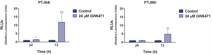
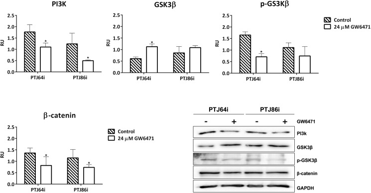
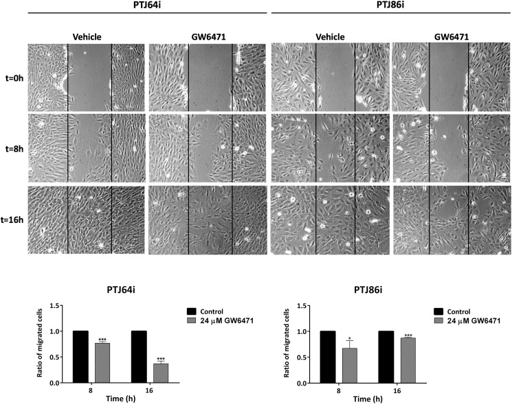
Head and neck paragangliomas (HNPGLs) are rare tumors that may cause important morbidity, because of their tendency to infiltrate the skull base. At present, surgery is the only therapeutic option, but radical removal may be difficult or impossible. Thus, effective targets and molecules for HNPGL treatment need to be identified. However, the lack of cellular models for this rare tumor hampers this task. PPARα receptor activation was reported in several tumors and this receptor appears to be a promising therapeutic target in different malignancies. Considering that the role of PPARα in HNPGLs was never studied before, we analyzed the potential of modulating PPARα in a unique model of HNPGL cells. We observed an intense immunoreactivity for PPARα in HNPGL tumors, suggesting that this receptor has an important role in HNPGL. A pronounced nuclear expression of PPARα was also confirmed in HNPGL-derived cells. The specific PPARα agonist WY14643 had no effect on HNPGL cell viability, whereas the specific PPARα antagonist GW6471 reduced HNPGL cell viability and growth by inducing cell cycle arrest and caspase-dependent apoptosis. GW6471 treatment was associated with a marked decrease of CDK4, cyclin D3 and cyclin B1 protein expression, along with an increased expression of p21 in HNPGL cells. Moreover, GW6471 drastically impaired clonogenic activity of HNPGL cells, with a less marked effect on cell migration. Notably, the effects of GW6471 on HNPGL cells were associated with the inhibition of the PI3K/GSK3β/β-catenin signaling pathway. In conclusion, the PPARα antagonist GW6471 reduces HNPGL cell viability, interfering with cell cycle and inducing apoptosis. The mechanisms affecting HNPGL cell viability involve repression of the PI3K/GSK3β/β-catenin pathway. Therefore, PPARα could represent a novel therapeutic target for HNPGL.


Conflict of interest statement

Competing Interests: The authors have declared that no competing interests exist.

Figures

Fig 1
Fig 1. Immunohistochemical analysis of PPARα in HNPGL tissues.
Tumor sections derived from two different HNPGL patients show positive immunostaining for PPARα in paraganglioma cell clusters, while the supporting stroma is negative. Bar = 80 μm.
Fig 2
Fig 2. Immunofluorescence analysis of PPARs in HNPGL cells.
PTJ64i (left panels) and PTJ86i (right panels) show high levels of PPARα nuclear expression. The fluorescence intensity for PPARα is stronger as compared to that for PPARβ/δ or PPARγ. Bar = 20 μm.
Fig 3
Fig 3. Effect of the PPARα agonist WY14643, or antagonist GW6471 on cell viability in PTJ64i and PTJ86i cell lines.
Cells were incubated for 72 hours with WY14643 or GW6471 at the indicated concentrations, or with 0.16% DMSO vehicle (control). GW6471 significantly inhibited viability in both cell lines (IC50 of 10 μM in PTJ64i and 16 μM in PTJ86i). Data shown are the means ±SD of three experiments with quintuplicate determinations. *Significant differences between control and each drug concentration (*p<0.05; **p<0.01; ***p<0.001).
Fig 4
Fig 4. Growth curves of PTJ64i and PTJ86i cells treated with GW6471.
Cell number was measured over a 72-hour time course treatment with 24 μM GW6471 or with vehicle control. Data shown are the means ±SD of three to five determinations (*p<0.05; **p<0.01; ***p<0.001).
Fig 5
Fig 5. Effect of GW6471 on PTJ64i and PTJ86i cell cycles.
The histograms show the mean percentage of cells (values inside the bars) in the different cell phases after a 24-hour treatment with 24 μM GW6471 as compared to control (**p<0.01; ***p<0.001).
Fig 6
Fig 6. Effect of GW6471 on the expression of relevant cell cycle proteins in PTJ64i and PTJ86i cell lines.
Expression of relevant cell cycle proteins after incubation of cells for 24 hours with 24 μM GW6471 was analyzed by Western blot using antibodies directed against the indicated proteins.
Fig 7
Fig 7. Apoptosis in PTJ64i and PTJ86i cells treated with 24 μM GW6471 for 24, 48 or 72 hours.
Values represented in the histograms (top) are the means ±SD of at least two independent determinations (*p<0.05; **p<0.01; ***p<0.001). Dot plots (bottom) show representative experiments after a 72-hour treatment with 24 μM GW6471.
Fig 8
Fig 8. Caspase 3/7 activity in PTJ64i and PTJ86i cells treated with 24 μM GW6471 for 24 and 72 hours.
Values represented in the histograms are the means ±SD of six determinations (*p<0.05; **p<0.01; ***p<0.001).
Fig 9
Fig 9. Effect of GW6471 on PI3K/GSK-3β/β-catenin signaling pathway in PTJ64i and PTJ86i cell lines.
Histograms of normalized densitometric analyses and representative western blotting for PI3K, GSK3β, p-GSK3β and β-catenin in HNPGL cells treated with 24 μM GW6471 for 72 hours are shown. The relative densities of the immunoreactive bands were determined and normalized with respect to GAPDH (loading control) using a semiquantitative densitometric analysis (Kodak ID Image Analysis Software, Rochester, NY, USA). Values are expressed as relative units (RU). Each bar represents the mean ± SD of two up to five independent determinations (*p<0.05).
Fig 10
Fig 10. Effect of GW6471 on clonogenic activity of PTJ64i and PTJ86i cell lines.
Data shown in the histograms are the means ±SD of three independent experiments (*p<0.05; **p<0.01). PE: plating efficiency [(# of colonies formed/# of cells plated)*100]; SF: surviving fraction [# of colonies formed *100/(# of cells plated *PE of control vehicle)].
Fig 11
Fig 11. Wound healing assay.
The effect of 24 μM GW6471 on HNPGL cell migration was evaluated by a monolayer wound-healing assay. Pictures of PTj64i and PTJ86i cells treated with vehicle or with GW6471 were taken at 0, 8 and 16 hours to analyze the dynamics of wound closure (vertical lines indicate wound edges). Histograms represent quantitative analyses of cell migration and are expressed as the ratio of the number of migrated cells in three fields after treatment as compared with vehicle (*p<0.05; ***p<0.001).

References

    1. Taïeb D, Kaliski A, Boedeker CC, Martucci V, Fojo T, Adler JR Jr, et al. Current approaches and recent developments in the management of head and neck paragangliomas. Endocr Rev. 2014. October;35: 795–819. doi: 10.1210/er.2014-1026 - DOI - PMC - PubMed
    1. Baysal BE, Ferrell RE, Willett-Brozick JE, Lawrence EC, Myssiorek D, Bosch A, et al. Mutations in SDHD, a mitochondrial complex II gene, in hereditary paraganglioma. Science. 2000. February 4;287: 848–851. - PubMed
    1. Rustin P, Munnich A, Rötig A. Succinate dehydrogenase and human diseases: new insights into a well-known enzyme. Eur J Hum Genet. 2002. May;10: 289–291. doi: 10.1038/sj.ejhg.5200793 - DOI - PubMed
    1. Lack EE, Lloyd RV, Carney JA, Woodruff JM; Association of Directors of Anatomic and Surgical Pathology. Recommendations for reporting of extra-adrenal paragangliomas. Mod Pathol. 2003. August;16: 833–855. doi: 10.1097/01.MP.0000081050.89276.CA - DOI - PubMed
    1. Fliedner SM, Lehnert H, Pacak K. Metastatic paraganglioma. Semin Oncol. 2010. December;37: 627–637. doi: 10.1053/j.seminoncol.2010.10.017 - DOI - PMC - PubMed

MeSH terms