. 2017 Jul 17;30(7):1406-1418.
doi: 10.1021/acs.chemrestox.7b00052. Epub 2017 Jun 26.

Diacetyl/l-Xylulose Reductase Mediates Chemical Redox Cycling in Lung Epithelial Cells

Affiliations

Diacetyl/l-Xylulose Reductase Mediates Chemical Redox Cycling in Lung Epithelial Cells

Shaojun Yang et al. Chem Res Toxicol. .

Abstract

Reactive carbonyls such as diacetyl (2,3-butanedione) and 2,3-pentanedione in tobacco and many food and consumer products are known to cause severe respiratory diseases. Many of these chemicals are detoxified by carbonyl reductases in the lung, in particular, dicarbonyl/l-xylulose reductase (DCXR), a multifunctional enzyme important in glucose metabolism. DCXR is a member of the short-chain dehydrogenase/reductase (SDR) superfamily. Using recombinant human enzyme, we discovered that DCXR mediates redox cycling of a variety of quinones generating superoxide anion, hydrogen peroxide, and, in the presence of transition metals, hydroxyl radicals. Redox cycling activity preferentially utilized NADH as a cosubstrate and was greatest for 9,10-phenanthrenequinone and 1,2-naphthoquinone, followed by 1,4-naphthoquinone and 2-methyl-1,4-naphthoquinone (menadione). Using 9,10-phenanthrenequinone as the substrate, quinone redox cycling was found to inhibit DCXR reduction of l-xylulose and diacetyl. Competitive inhibition of enzyme activity by the quinone was observed with respect to diacetyl (Ki = 190 μM) and l-xylulose (Ki = 940 μM). Abundant DCXR activity was identified in A549 lung epithelial cells when diacetyl was used as a substrate. Quinones inhibited reduction of this dicarbonyl, causing an accumulation of diacetyl in the cells and culture medium and a decrease in acetoin, the reduced product of diacetyl. The identification of DCXR as an enzyme activity mediating chemical redox cycling suggests that it may be important in generating cytotoxic reactive oxygen species in the lung. These activities, together with the inhibition of dicarbonyl/l-xylulose metabolism by redox-active chemicals, as well as consequent deficiencies in pentose metabolism, are likely to contribute to lung injury following exposure to dicarbonyls and quinones.


Conflict of interest statement

The authors declare no competing financial interest

Figures

Figure 1
Figure 1
Enzymatic reactions of DCXR. DCXR can mediate the two electron NAD(P)H-dependent reduction of typical substrates including diacetyl and L-xylulose. In the absence of these substrates, DCXR can also mediate the NAD(P)H-dependent one electron reduction of quinones forming semiquinones. In this reaction, NAD(P)H generates two molecules of the semiquinone. Reaction of the semiquinones with molecular oxygen regenerates the parent compound forming a superoxide anion in the process. It should be noted that it is possible that DCXR also mediates the NAD(P)H-dependent two electron reduction of quinones to the hydroquinone. Autooxidation of the hydroquinone can result in the one electron reduced quinone, which can then react with oxygen in the redox cycling process.
Figure 2
Figure 2
Purification and activity of recombinant human DCXR. (Panel A) SDS–PAGE analysis of recombinant human DCXR expressed in E. coli, lane M, protein standard. Lane 1, crude extract of E. coli containing DCXR induced with 0.5 mM isopropyl-D-thiogalactopyranoside; lane 2, flow-through fraction from the Ni-NTA column; lane 3, imidazole eluted fraction from nickel-affinity column. The arrow indicates the purified enzyme. (Panel B) Effects of increasing concentrations of DCXR on reduction of 5 mM diacetyl as measured by NADH oxidation. Reactions contained 600 μM NADH. (Inset) Effects of increasing DCXR protein in reaction mixes on rates of diacetyl reduction. (Panels C and D) Comparison of NADH and NADPH in supporting diacetyl or L-xylulose reduction by DCXR. Reaction mixes contained 200 μM NADH or NADPH and enzyme activity measured by changes in absorbance at 340 nm. (Panel E) Comparison of H2O2 generation by DCXR in reaction containing NADH or NADPH in the presence of 20 μM 9,10-phenanthrenequinone. Data are presented as the mean ± SE (n = 4), significantly more DCXR activity was evident in all reactions containing NADH when compared to NADPH (p 0.001, from t test analysis using Sigmaplot).
Figure 3
Figure 3
Generation of reactive oxygen species by human recombinant DCXR. (Panel A) Effects of menadione on superoxide anion production by DCXR in the presence of 600 μM NADH. Menadione (300 μM) was added to reaction mixes as indicated by the arrow to stimulate superoxide anion formation. Reaction mixes in 100 μL contained 2.56 μg DCXR. Superoxide dismutase (SOD) (400 U/mL) was added to the reactions after 3.5 min as indicated by the arrowhead. Control reactions did not contain DCXR. (Panel B) Effects of menadione (300 μM) on H2O2 production by DCXR. Reactions were run in the presence and absence of catalase (2 kU/mL). Menadione was added at time zero. Reaction mixes in 100 μL contained 1.93 μg of DCXR. (Panel C) Effects of 9,10-phenanthrenequinone (9,10-PQ) on the production of hydroxyl radicals by DCXR. Reactions were run in the presence and absence of DMSO (15 mM). 9,10-Pheneanthrenequinone (20 μM) was added at time zero. Reaction mixes in 100 μL contained 1.6 μg of DCXR and 2 mM terepthalate. (Inset) Ability of hydroxyl radicals generated by DCXR redox cycling to nick closed circular plasmid DNA. All samples in 20 μL reaction mixes contained 200 ng of plasmid DNA and 20 μM 9,10-phenanthrenequinone. Reactions were run in the absence (lane 1) and presence of DCXR (lanes 2–5). Samples in lanes 3 and 5 contained 100 μM Fe2+ and 110 μM EDTA; samples in lanes 4 and 5 contained 100 U of catalase. Reaction mixes were incubated for 1 h and then analyzed on a 0.8% agarose gel. Closed circular plasmid DNA and nicked plasmid DNA bands were visualized under ultraviolet light and are indicated by arrowhead and arrow, respectively. (Panel D) Ability of different quinones to redox cycling with DCXR. H2O2 formation was measured in the presence of 200 μM NADPH and increasing concentrations of redox cycling chemicals. The maximum values of H2O2 production by DCXR for each quinones are listed in Table 2.
Figure 4
Figure 4
Oxygen consumption by human recombinant DCXR during chemical redox cycling. The reaction was initially run in the presence of NADH (600 μM) and 9,10-phenanthrenequinone (20 μM) to establish a stable baseline. DCXR was then added to start the reaction. (Panel A) Inhibition of oxygen consumption by diacetyl (5 mM) which was added after DCXR (arrow). (Panel B) Pretreatment with diacetyl inhibited oxygen consumption during redox cycling by DCXR. Diacetyl was added to the reaction mix at time zero; 9,10-phenanthrenequinone (9,10-PQ) was used as the redox cycling chemical. (Panels C and D) Inhibition of oxygen consumption by hexanoic acid (50 mM) and butyric acid (10 mM) during redox cycling by DCXR. The short chain fatty acids were added at the time points indicated.
Figure 5
Figure 5
Redox cyclers inhibit acetoin formation during diacetyl reduction by recombinant human DCXR. (Panel A) Effects of menadione on diacetyl reduction by DCXR. Reaction mixes contained 2 mM diacetyl, 600 μM NADH, and 10 μg/mL DCXR. At the indicated times, aliquots were removed and analyzed by HPLC for diacetyl and acetoin as described in the Materials and Methods. The top HPLC tracing shows the acetoin standard (10 nmol). The second and third HPLC tracings show diacetyl metabolism in reaction mixes after 0 and 10 min, respectively. The lower HPLC tracing shows the effects of menadione (300 μM) on the reduction of diacetyl. (Panel A, inset) In the presence of 300 μM menadione (MD), > 90% of the DCXR mediated reduction of diacetyl was inhibited. (Panel B) Effects of redox cyclers on diacetyl reduction by recombinant DCXR. Enzyme activity was assayed by the formation of acetoin in reaction mixes by HPLC. IC50 values for 1,2-naphthoquinone (1,2-NQ), 9,10-phenanthrenequinone (9,10-PQ), 1,4-naphthoquinone (1,4-NQ), and menadione (MD) are 2.9, 9.3, 11.8, and 52.8 μM, respectively. (Panel C) Lineweaver–Burk plot showing a competitive type of inhibition of diacetyl reduction by menadione.
Figure 6
Figure 6
Effects of diacetyl and quinones on DCXR. (Panel A) Diacetyl restores DCXR substrate reduction inhibited by menadione (MD). DCXR reactions were run in the absence (gray bars) and presence (black bars) of 300 μM menadione, 200 μM NADPH, and 5 mM diacetyl. After 0, 5, or 10 min, substrates and cofactors were removed using Histag-agarose beads. Imidazole (200 mM) was used to elute DCXR from the beads, and then 10 μL of eluent fraction was added to 90 μL of enzyme reaction mixes which contained 200 μM NADPH and 5 mM diacetyl. Imidazole (20 mM) in the final reaction mix did not affect enzyme activity. Lane a is a positive control. In lane b, menadione was added back to the reaction mixtures where indicated. In lanes c–h, no menadione was added back to the reaction mixtures. DCXR activity was then measured over an additional 10 min incubation time. Reduction of diacetyl to acetoin was measured by HPLC as described in the Materials and Methods. These data demonstrate that DCXR-mediated inhibition of diacetyl reduction by menadione was reversible. (Panel B) Diacetyl (5 mM) inhibits superoxide anion formation by DCXR in the presence of 300 μM menadione. Reaction mixes in 100 μL contained 1.6 μg of DCXR and 600 μM NADH. (Inset) In the presence of diacetyl, menadione-stimulated superoxide anion formation by DCXR was inhibited by approximately 75%. Data are presented as the mean ± SE (n = 3). (Panels C and D) Lineweaver–Burk plots showing competitive type inhibition of chemical redox cycling by diacetyl and L-xylulose, respectively, of cytochrome c reduction induced by 9,10-phenanthrenequinone. Reaction mixes in 100 μL contained either 1.6 μg or 1.2 μg of DCXR in panels B and C, respectively.
Figure 7
Figure 7
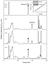
Metabolism of diacetyl by lung epithelial cells. A549 cells were treated with diacetyl (2 mM) as described in Materials and Methods. After 0, 2, and 3 h, the culture medium was assayed for diacetyl and acetoin. The upper HPLC tracing shows an acetoin standard. The lower HPLC tracings show acetoin accumulation in the culture medium after 0, 2, and 3 h. An additional peak at about 20 min was generated in the reaction. This peak likely responds to another metabolite of diacetyl. (Inset) Western blot showing the expression of DCXR in A549 cells from two independent samples and quantification of acetoin accumulation in the culture medium of A549 cells after the addition of diacetyl. In the Western blot, β-actin was used as a protein loading control.
Figure 8
Figure 8
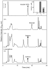
Effects of menadione on the metabolism of diacetyl in lung epithelial cells. A549 cells were treated with diacetyl (2 mM) in the absence and presence of menadione (MD, 300 μM). The upper HPLC tracing shows an acetoin standard. The center two HPLC tracings show acetoin accumulation in the culture medium at 0 and 3 h after the addition of diacetyl. The lower HPLC tracing shows that acetoin formation in the cells is inhibited in the presence of menadione. (Inset) After 3 h, menadione inhibits diacetyl metabolism by approximately 65%.
Figure 9
Figure 9
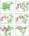
Molecular docking of L-xylulose, diacetyl, and redox cycling quinones to human DCXR. The docking model of substrates or redox cycler bound to human DCXR (PDB ID: 1WNT) was performed using AutoDock Vina; the best-ranked docking poses were analyzed using PyMOL. (A,B) Docking of L-xylulose to human DCXR. The crystal structure of DCXR is shown as a ribbon diagram in green with critical amino acids (including catalytic triad S136, Y149, and K153) depicted as sticks with green backbones (oxygen, red; nitrogen, blue; and phosphor, orange). NADP is shown as sticks with magenta backbones. L-Xylulose is shown in spherical form (A) or in stick form (B) with cyan backbones. Residues within 4 Å of L-xylulose are indicated. Hydrogen bonds are presented in red broken lines, and the corresponding distances (Å) are indicated. The best-ranked docking pose of L-xylulose is situated in proximity to the nicotinamide moiety of NADP and the catalytic residues (S136 and Y149) of DCXR. L-Xylulose forms H bond contacts with Y149, H146, W191, and the oxygen atom on the nicotinamide moiety of NADP. (C) Interactions of diacetyl with key amino acid residues in DCXR. Diacetyl, shown in sticks with gray backbones, is located within van der Waals contacts with Y149, L89, and V181 and forms H bond interactions with H146 and W191. (D) Interactions of 1,4-naphthoquinone with key amino acid residues in DCXR. 1,4-Naphthoquinone, shown as sticks with yellow backbones, fits in the hydrophobic cleft in the active site of DCXR forming hydrophobic interactions with M186, V181, W191, H146, L89, and Y149. (E) Superposition of L-xylulose, diacetyl, and 1,4-naphthoquinone in human DCXR. Note that the docked positions of L-xylulose, diacetyl, and 1,4-naphthoquinone are highly similar in the active site of DCXR, suggesting that these compounds may act as competitive substrates/inhibitors of the enzyme. (F) Superposition of diacetyl and 9,10-phenanthrenequinone in human DCXR. 9,10-Phenanthrenequinone is shown as sticks with orange backbones.

Similar articles

Cited by

References

    1. Ebert B, Kisiela M, Maser E. Human DCXR - another ’moonlighting protein’ involved in sugar metabolism, carbonyl detoxification, cell adhesion and male fertility? Biol Rev Camb Philos Soc. 2015;90:254–278. - PubMed
    1. Nakagawa J, Ishikura S, Asami J, Isaji T, Usami N, Hara A, Sakurai T, Tsuritani K, Oda K, Takahashi M, Yoshimoto M, Otsuka N, Kitamura K. Molecular characterization of mammalian dicarbonyl/l-xylulose reductase and its localization in kidney. J Biol Chem. 2002;277:17883–17891. - PubMed
    1. Asami J, Odani H, Ishii A, Oide K, Sudo T, Nakamura A, Miyata N, Otsuka N, Maeda K, Nakagawa J. Suppression of AGE precursor formation following unilateral ureteral obstruction in mouse kidneys by transgenic expression of alpha-dicarbonyl/l-xylulose reductase. Biosci Biotechnol, Biochem. 2006;70:2899–2905. - PubMed
    1. Touster O, Hutcheson RM, Rice L. The influence of D-glucuronolactone on the excretion of l-xylulose by humans and guinea pigs. Biochim Biophys Acta. 1955;215:677–684. - PubMed
    1. Winegrad AI, Burden CL. l-xylulose metabolism in diabetes mellitus. N Engl J Med. 1966;274:298–305. - PubMed

Publication types

LinkOut - more resources