

Genetic Lineage Tracing in Taste Tissues Using Sox2-CreERT2 Strain
- PMID: 28595328
- PMCID: PMC6075561
- DOI: 10.1093/chemse/bjx032
Genetic Lineage Tracing in Taste Tissues Using Sox2-CreERT2 Strain
Abstract
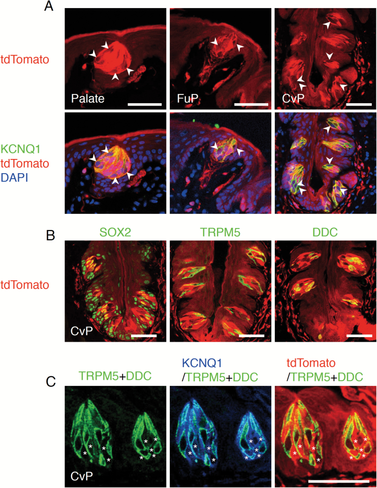
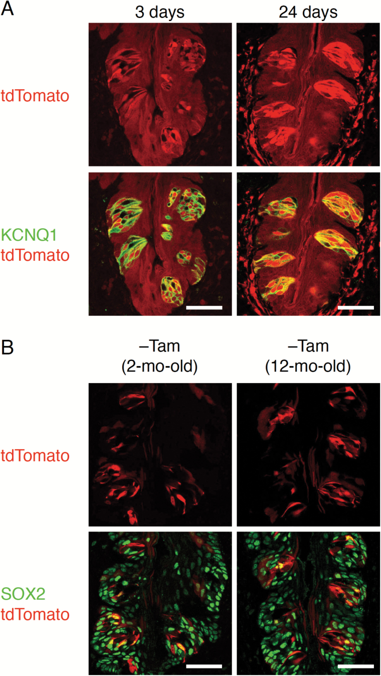
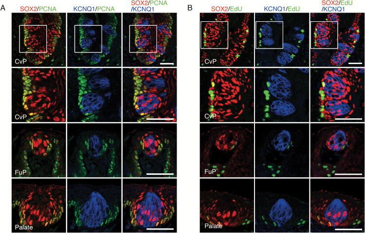
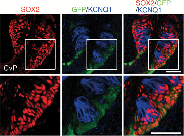
Taste cells in taste buds are epithelial sensory cells. Old taste bud cells die and are replaced by new ones generated from taste stem cells. Identifying and characterizing adult taste stem cells is therefore important to understand how peripheral taste tissues are maintained. SOX2 is expressed in oral epithelium including gustatory papillae and has been proposed to be a marker of adult taste stem/progenitor cells. Nevertheless, this hypothesis has never been directly tested. Here, by single-color genetic lineage tracing using Sox2-CreERT2 strain, we reveal that all types of taste bud cells distributed throughout the oral epithelium are derived from stem cells that express SOX2. Short-term tracing shows that SOX2-positive taste stem cells actively supply taste bud cells. At the base of epithelium outside taste buds are distributed proliferation marker- and SOX2-positive cells. Consistently, taste stem cells identified by Lgr5 expression in the circumvallate papillae also express SOX2. Together, taste stem cells distributed in oral epithelia express SOX2.
Keywords: lineage tracing; oral epithelium; stem cell; taste bud cell.
© The Author 2017. Published by Oxford University Press. All rights reserved. For permissions, please e-mail: journals.permissions@oup.com.
Figures
Similar articles
-
SOX2 regulates homeostasis of taste bud cells and lingual epithelial cells in posterior tongue.PLoS One. 2020 Oct 15;15(10):e0240848. doi: 10.1371/journal.pone.0240848. eCollection 2020. PLoS One. 2020. PMID: 33057384 Free PMC article.
-
Maintenance and turnover of Sox2+ adult stem cells in the gustatory epithelium.PLoS One. 2022 Sep 2;17(9):e0267683. doi: 10.1371/journal.pone.0267683. eCollection 2022. PLoS One. 2022. PMID: 36054203 Free PMC article.
-
Single Lgr5- or Lgr6-expressing taste stem/progenitor cells generate taste bud cells ex vivo.Proc Natl Acad Sci U S A. 2014 Nov 18;111(46):16401-6. doi: 10.1073/pnas.1409064111. Epub 2014 Nov 3. Proc Natl Acad Sci U S A. 2014. PMID: 25368147 Free PMC article.
-
How taste works: cells, receptors and gustatory perception.Cell Mol Biol Lett. 2015 Dec;20(5):699-716. doi: 10.1515/cmble-2015-0042. Cell Mol Biol Lett. 2015. PMID: 26447485 Review.
-
In vivo fate tracing studies of mammalian taste cell progenitors.Ann N Y Acad Sci. 2009 Jul;1170:34-8. doi: 10.1111/j.1749-6632.2009.04371.x. Ann N Y Acad Sci. 2009. PMID: 19686103 Free PMC article. Review.
Cited by
-
The sense of taste: Development, regeneration, and dysfunction.WIREs Mech Dis. 2022 May;14(3):e1547. doi: 10.1002/wsbm.1547. Epub 2021 Nov 30. WIREs Mech Dis. 2022. PMID: 34850604 Free PMC article. Review.
-
EGR4 is critical for cell-fate determination and phenotypic maintenance of geniculate ganglion neurons underlying sweet and umami taste.Proc Natl Acad Sci U S A. 2023 May 30;120(22):e2217595120. doi: 10.1073/pnas.2217595120. Epub 2023 May 22. Proc Natl Acad Sci U S A. 2023. PMID: 37216536 Free PMC article.
-
Early Steps towards Hearing: Placodes and Sensory Development.Int J Mol Sci. 2023 Apr 10;24(8):6994. doi: 10.3390/ijms24086994. Int J Mol Sci. 2023. PMID: 37108158 Free PMC article. Review.
-
Cellular Diversity and Regeneration in Taste Buds.Curr Opin Physiol. 2021 Apr;20:146-153. doi: 10.1016/j.cophys.2021.01.003. Epub 2021 Jan 12. Curr Opin Physiol. 2021. PMID: 33615087 Free PMC article.
-
SOX2 regulates homeostasis of taste bud cells and lingual epithelial cells in posterior tongue.PLoS One. 2020 Oct 15;15(10):e0240848. doi: 10.1371/journal.pone.0240848. eCollection 2020. PLoS One. 2020. PMID: 33057384 Free PMC article.
References
-
- Asano-Miyoshi M, Hamamichi R, Emori Y. 2008. Cytokeratin 14 is expressed in immature cells in rat taste buds. J Mol Histol. 39:193–199. - PubMed
-
- Barker N, Huch M, Kujala P, van de Wetering M, Snippert HJ, van Es JH, Sato T, Stange DE, Begthel H, van den Born M et al. . 2010. Lgr5(+ve) stem cells drive self-renewal in the stomach and build long-lived gastric units in vitro. Cell Stem Cell. 6:25–36. - PubMed
-
- Barker N, van Es JH, Kuipers J, Kujala P, van den Born M, Cozijnsen M, Haegebarth A, Korving J, Begthel H, Peters PJ et al. . 2007. Identification of stem cells in small intestine and colon by marker gene Lgr5. Nature. 449:1003–1007. - PubMed
-
- Barker N, van Oudenaarden A, Clevers H. 2012. Identifying the stem cell of the intestinal crypt: strategies and pitfalls. Cell Stem Cell. 11:452–460. - PubMed
MeSH terms
Substances
Grants and funding
LinkOut - more resources
Full Text Sources
Other Literature Sources
