Review
. 2017 Jun 8;10(1):287.
doi: 10.1186/s13071-017-2204-7.

The evolution of trypanosomatid taxonomy

Affiliations
Review

The evolution of trypanosomatid taxonomy

Alexa Kaufer et al. Parasit Vectors. .

Abstract

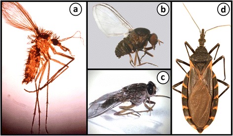
Trypanosomatids are protozoan parasites of the class Kinetoplastida predominately restricted to invertebrate hosts (i.e. possess a monoxenous life-cycle). However, several genera are pathogenic to humans, animals and plants, and have an invertebrate vector that facilitates their transmission (i.e. possess a dixenous life-cycle). Phytomonas is one dixenous genus that includes several plant pathogens transmitted by phytophagous insects. Trypanosoma and Leishmania are dixenous genera that infect vertebrates, including humans, and are transmitted by hematophagous invertebrates. Traditionally, monoxenous trypanosomatids such as Leptomonas were distinguished from morphologically similar dixenous species based on their restriction to an invertebrate host. Nonetheless, this criterion is somewhat flawed as exemplified by Leptomonas seymouri which reportedly infects vertebrates opportunistically. Similarly, Novymonas and Zelonia are presumably monoxenous genera yet sit comfortably in the dixenous clade occupied by Leishmania. The isolation of Leishmania macropodum from a biting midge (Forcipomyia spp.) rather than a phlebotomine sand fly calls into question the exclusivity of the Leishmania-sand fly relationship, and its suitability for defining the Leishmania genus. It is now accepted that classic genus-defining characteristics based on parasite morphology and host range are insufficient to form the sole basis of trypanosomatid taxonomy as this has led to several instances of paraphyly. While improvements have been made, resolution of evolutionary relationships within the Trypanosomatidae is confounded by our incomplete knowledge of its true diversity. The known trypanosomatids probably represent a fraction of those that exist and isolation of new species will help resolve relationships in this group with greater accuracy. This review incites a dialogue on how our understanding of the relationships between certain trypanosomatids has shifted, and discusses new knowledge that informs the present taxonomy of these important parasites.

Keywords: Leishmania; Leptomonas; Phylogenetics; Systematics; Taxonomy; Trypanosomatid; Zelonia.


Figures

Fig. 1
Fig. 1
Vectors and invertebrate hosts of some trypanosomatids. a A female Phlebotomus sp. sand fly which is a vector of Leishmania spp. Citation: Hailu et al. Visceral leishmaniasis: New health tools are needed. PLoS Med. 2005;2(7):590–594 [151]. b A female Simulium (Morops) dycei, which is the host of Zelonia australiensis. Citation: Barratt et al. Isolation of novel trypanosomatid, Zelonia australiensis sp. nov. (Kinetoplastida: Trypanosomatidae) provides support for a Gondwanan origin of dixenous parasitism in the Leishmaniinae. PLOS Negl Trop Dis. 2017;11(1):e0005215 [68]. c The tsetse fly is the vector of Trypanosoma brucei; the aetiological agent of Human African Trypanosomiasis (http://researchnews.plos.org/2016/08/08/under-my-skin/) [152]. d A Triatomine “kissing” bug, which is the natural vector of Trypanosoma cruzi; the aetiological agent of Chagas disease. Citation: Curtis-Robles et al. Combining public health education and disease ecology research: using citizen science to assess Chagas disease entomological risk in Texas. PLoS Neglect Trop Dis. 2015;9(12):12 [153]. a, b, d Copyright: Creative Commons Attribution 4.0 International License (https://creativecommons.org/licenses/by/4.0/)
Fig. 2
Fig. 2
The six major morphotype classes of trypanosomatids. a Trypomastigote. b Epimastigote. c Amastigote. d Opisthomastigote. e Choanomastigote. f Promastigote. Forms a and b represent the juxtaform superclass and possess a flagellum that is laterally attached to the cell body. Forms d, e and f represent the liberform superclass and do not possess a laterally attached flagellum. Amastigotes (c) exist for both liberform and juxtaform trypanosomatids
Fig. 3
Fig. 3
Some clinical manifestations of leishmaniasis. a A patient with mucocutaneous leishmaniasis (MCL) presenting with facial ulcerative lesions and nasal obstruction. Cropped from original. Citation: Gois et al. Immune response to Leishmania antigens in an AIDS patient with mucocutaneous leishmaniasis as a manifestation of immune reconstitution inflammatory syndrome (IRIS): a case report. BMC Infect Dis. 2015;15(1):38 [154]. b Presentation of MCL with patients suffering from erythematous papules and ulcerations on the lip region. Cropped from original. Citation: Mohammadpour et al. Lip leishmaniasis: a case series with molecular identification and literature review. BMC Infect Dis. 2017;17(1) [155]. c A patient with cutaneous leishmaniasis presenting with crusted nodules over the left cheek (upper panel) and erythematous ulcerated plaques with crusts over the feet (lower panel). Cropped from original. Citation: Al-Dwibe et al. Contact dermatitis-like cutaneous leishmaniasis in a Libyan HIV patient. Parasit Vectors. 2014;7:3 [156]. a-c [157]
Fig. 4
Fig. 4
Photomicrographs of stained smears showing Leishmania infections. a May-Grunwald-Giemsa stained preparation from a case of feline leishmaniasis showing macrophages infected with L. infantum amastigotes. Cropped from original. Citation: Pennisi et al. [157]. LeishVet update and recommendations on feline leishmaniosis. Parasit Vectors. 2015;8(1):302. b Haematoxylin and eosin stained histological preparation from a canine deep dermis mucocutaneous lesion showing Leishmania amastigotes and Leishmania-infected fibroblasts (arrowheads). Cropped from original. Citation: Baneth et al. [158]. Mucocutaneous Leishmania tropica infection in a dog from a human cutaneous leishmaniasis focus. Parasit Vectors. 2014;7:5. c Haematoxylin and eosin stained preparation from a canine with cutaneous leishmaniasis showing intracellular Leishmania amastigotes in macrophages (arrows). Cropped from original. Citation: Ordeix et al. [159]. Histological and parasitological distinctive findings in clinically-lesioned and normal-looking skin of dogs with different clinical stages of leishmaniosis. Parasit Vectors. 2017;10:8. d May-Grunwald-Giemsa stained preparation from an aspirate of a mucocutaneous lesion predominately showing intracellular Leishmania amastigotes and few extracellular amastigotes. Cropped from original. Citation: Baneth et al. [158]. a-d Copyright: Creative Commons Attribution 4.0 International License (http://creativecommons.org/licenses/by/4.0/)
Fig. 5
Fig. 5
The growth cycle of trypanosomatids within invertebrates. a Replication of Leishmania (Leishmania) species in the sand fly vector occurs at two locations: procyclic promastigotes replicate in the abdominal midgut and leptomonad promastigotes in the thoracic midgut. The replicative procyclic promastigote forms differentiate into elongated nectomonad promastigotes that migrate anteriorly into the thoracic midgut, where further replication in the leptomonad form occurs. Some leptomonad promastigotes attach to the cuticle-lined surface of the midgut and differentiate into haptomonad promastigotes that may act as precursors for differentiation into metacyclic promastigotes, which is the stage infective to the mammalian host. b Leptomonas are ingested in the cyst form and differentiate into the promastigote form. In the crop, the leptomonad form undergoes fission and later in the midgut and pylorus by unequal fission or budding. Cysts are formed via budding in the rectum and are passed out in the faeces as the infective form
Fig. 6
Fig. 6
Diagrammatic representation of the three Leishmania sections proposed by Lainson & Shaw (1979). Figure shows the sections Hypopylaria, Suprapylaria and Peripylaria relative to the relevant structural features of the sand fly including the proboscis (pr), stomodeal valve (sv), cardia (c), thoracic midgut (tm), abdominal midgut (am), malpighian tubules (mt), pylorus (py) and rectum (r). The distribution of Leishmania development within the sand fly vector is shown in black
Fig. 7
Fig. 7
Light and electron micrographs of Zelonia australiensis. a Transmission electron micrograph showing the gross morphological features of Zelonia australiensis promastigotes including the nucleus (Nu), karysome (Ka), kinetoplast (K), flagella (fl), flagella pocket (fp), glycosomes (gl) and the Golgi body (gb). Subpelicular microtubules (S) give some cell edges a striated appearance, depending on the angle of sectioning. b, c Light micrographs showing promastigotes in a Leishman stained smear. d Light micrograph of a live-cell wet preparation viewed under phase contrast microscopy

Similar articles

Cited by

References

    1. Overath P, Haag J, Lischke A, O’HUigin C. The surface structure of trypanosomes in relation to their molecular phylogeny. Int J Parasitol. 2001;31(5–6):468–471. doi: 10.1016/S0020-7519(01)00152-7. - DOI - PubMed
    1. Bouteille B, Oukem O, Bisser S, Dumas M. Treatment perspectives for human African trypanosomiasis. Fundam Clin Pharmacol. 2003;17(2):171–181. doi: 10.1046/j.1472-8206.2003.00167.x. - DOI - PubMed
    1. Hemmige V, Tanowitz H, Sethi A. Trypanosoma cruzi infection: a review with emphasis on cutaneous manifestations. Int J Dermatol. 2012;51(5):501–508. doi: 10.1111/j.1365-4632.2011.05380.x. - DOI - PMC - PubMed
    1. Nussbaum K, Honek J, Cadmus C, Efferth T. Trypanosomatid parasites causing neglected diseases. Curr Med Chem. 2010;17(15):1594–1617. doi: 10.2174/092986710790979953. - DOI - PubMed
    1. Rassi A, Rassi A, de Rezende JM. American trypanosomiasis (Chagas disease) Infect Dis Clin North Am. 2012;26(2):275–291. doi: 10.1016/j.idc.2012.03.002. - DOI - PubMed

LinkOut - more resources