

HPV8 Field Cancerization in a Transgenic Mouse Model Is due to Lrig1+ Keratinocyte Stem Cell Expansion
- PMID: 28595997
- PMCID: PMC5613749
- DOI: 10.1016/j.jid.2017.04.039
HPV8 Field Cancerization in a Transgenic Mouse Model Is due to Lrig1+ Keratinocyte Stem Cell Expansion
Abstract
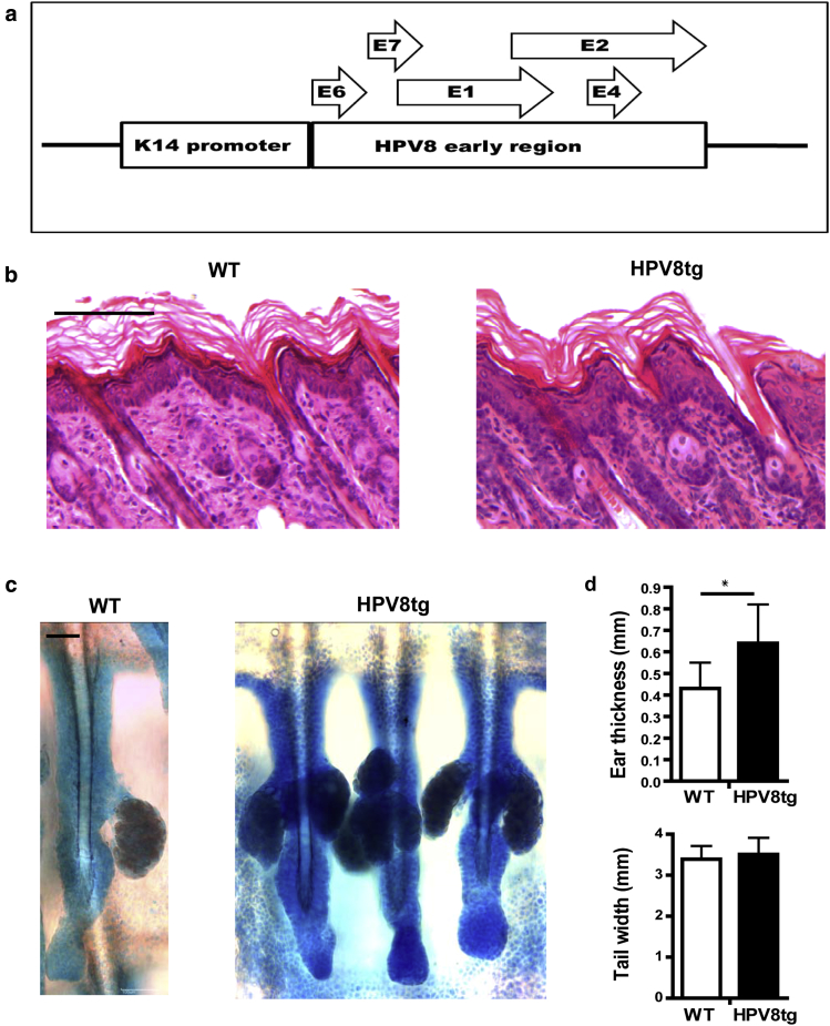
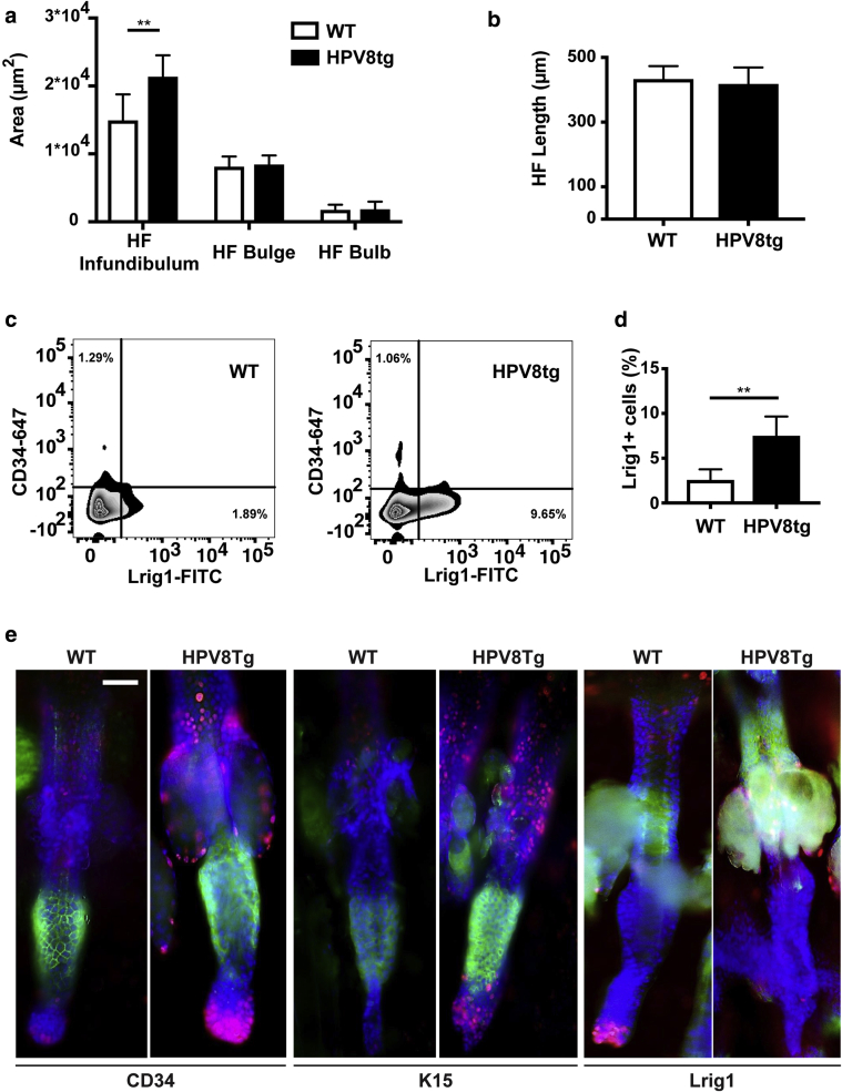
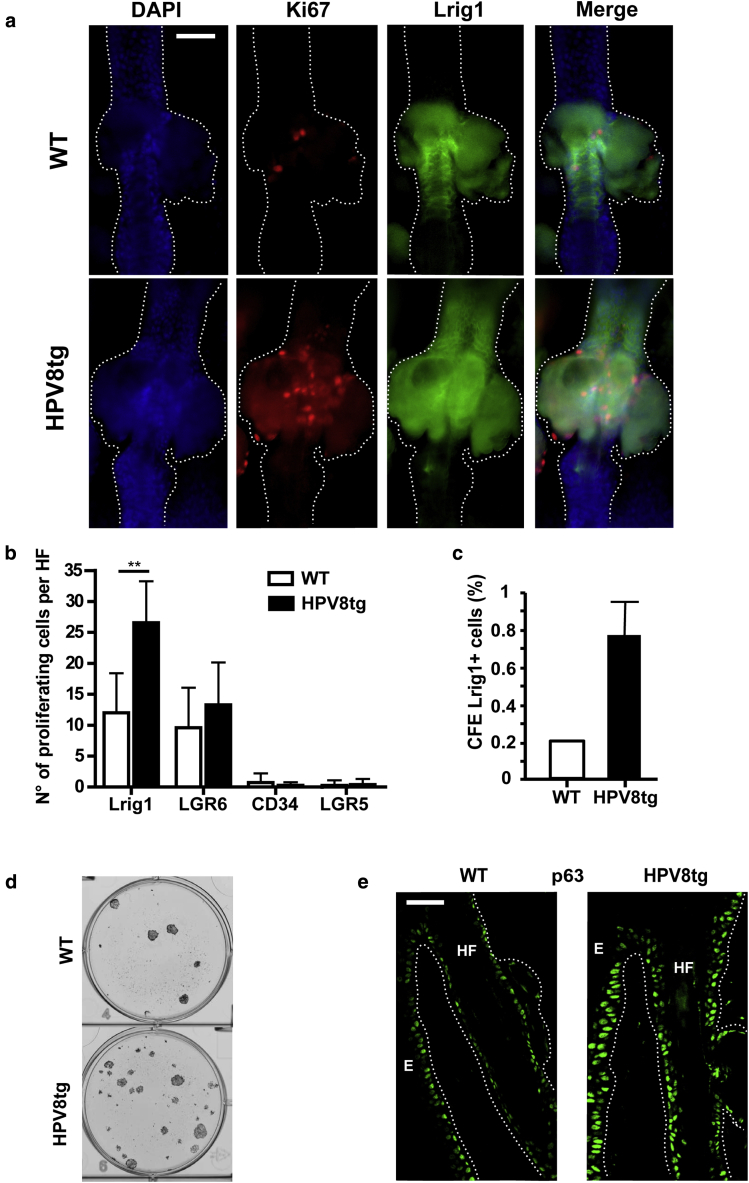
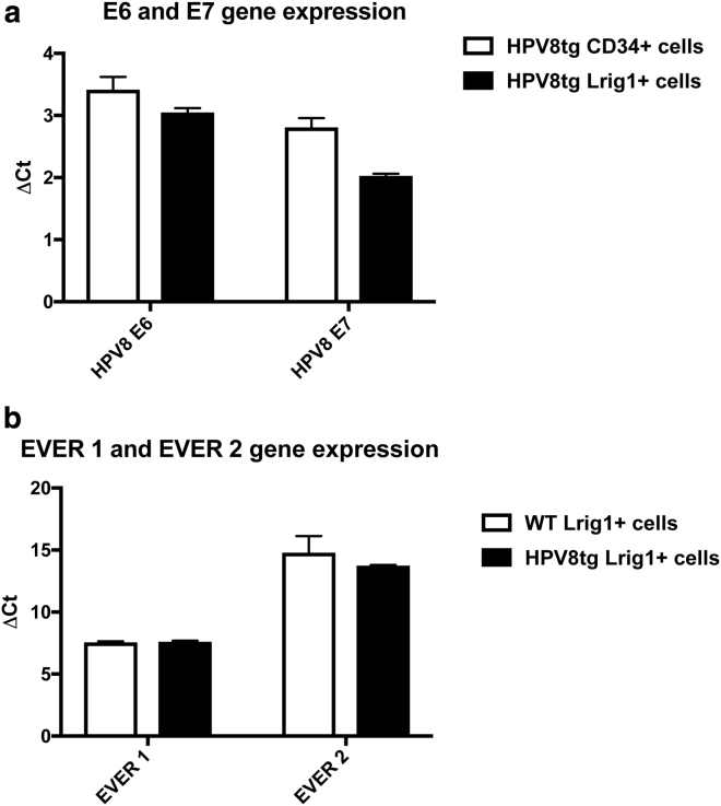
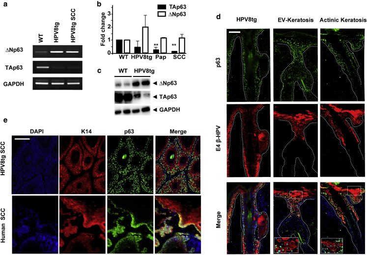
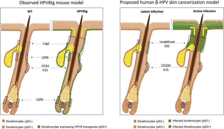
β-Human papillomaviruses (HPVs) cause near ubiquitous latent skin infection within long-lived hair follicle (HF) keratinocyte stem cells. In patients with epidermodysplasia verruciformis, β-HPV viral replication is associated with skin keratosis and cutaneous squamous cell carcinoma. To determine the role of HF keratinocyte stem cells in β-HPV-induced skin carcinogenesis, we utilized a transgenic mouse model in which the keratin 14 promoter drives expression of the entire HPV8 early region (HPV8tg). HPV8tg mice developed thicker skin in comparison with wild-type littermates consistent with a hyperproliferative epidermis. HF keratinocyte proliferation was evident within the Lrig1+ keratinocyte stem cell population (69 vs. 55%, P < 0.01, n = 7), and not in the CD34+, LGR5+, and LGR6+ keratinocyte stem cell populations. This was associated with a 2.8-fold expansion in Lrig1+ keratinocytes and 3.8-fold increased colony-forming efficiency. Consistent with this, we observed nuclear p63 expression throughout this population and the HF infundibulum and adjoining interfollicular epidermis, associated with a switch from p63 transcriptional activation isoforms to ΔNp63 isoforms in HPV8tg skin. Epidermodysplasia verruciformis keratosis and in some cases actinic keratoses demonstrated similar histology associated with β-HPV reactivation and nuclear p63 expression within the HF infundibulum and perifollicular epidermis. These findings would suggest that β-HPV field cancerization arises from the HF junctional zone and predispose to squamous cell carcinoma.
Copyright © 2017 The Authors. Published by Elsevier Inc. All rights reserved.
Figures
References
-
- Abbas O., Richards J.E., Yaar R., Mahalingam M. Stem cell markers (cytokeratin 15, cytokeratin 19 and p63) in in situ and invasive cutaneous epithelial lesions. Mod Pathol. 2011;24:90–97. - PubMed
-
- Azzimonti B., Mondini M., De Andrea M., Gioia D., Dianzani U., Mesturini R. CD8+ T-cell lymphocytopenia and lack of EVER mutations in a patient with clinically and virologically typical epidermodysplasia verruciformis. Arch Dermatol. 2005;141:1323–1325. - PubMed
-
- Banerjee M., Sarma N., Biswas R., Roy J., Mukherjee A., Giri A.K. DNA repair deficiency leads to susceptibility to develop arsenic-induced premalignant skin lesions. Int J Cancer. 2008;123:283–287. - PubMed
-
- Borgogna C., Landini M.M., Lanfredini S., Doorbar J., Bouwes Bavinck J.N., Quint K.D. Characterization of skin lesions induced by skin-tropic α- and β-papillomaviruses in a patient with epidermodysplasia verruciformis. Br J Dermatol. 2014;171:1550–1554. - PubMed
Publication types
MeSH terms
Substances
LinkOut - more resources
Full Text Sources
Other Literature Sources
Medical
Molecular Biology Databases
Research Materials
Miscellaneous
