Review
. 2017 Sep 10:261:62-86.
doi: 10.1016/j.jconrel.2017.05.033. Epub 2017 Jun 27.

Multifunctional nanodiamonds in regenerative medicine: Recent advances and future directions

Affiliations
Review

Multifunctional nanodiamonds in regenerative medicine: Recent advances and future directions

Jonathan Whitlow et al. J Control Release. .

Abstract

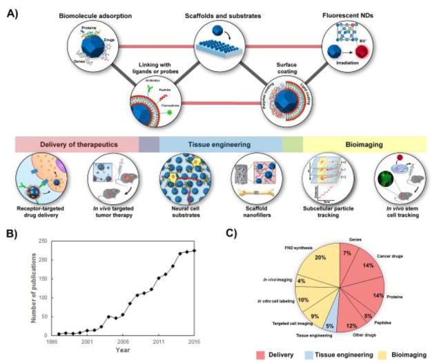
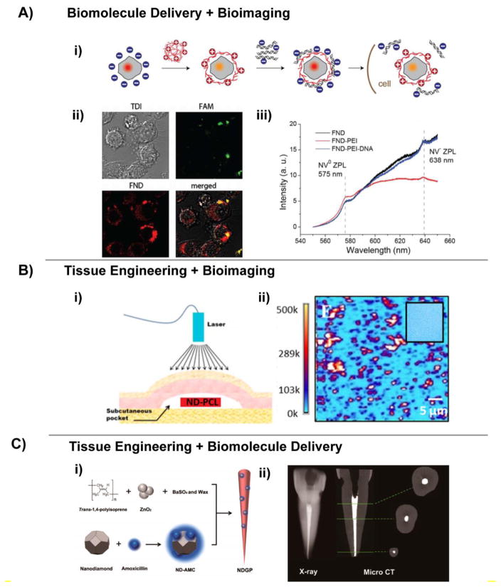
With recent advances in the field of nanomedicine, many new strategies have emerged for diagnosing and treating diseases. At the forefront of this multidisciplinary research, carbon nanomaterials have demonstrated unprecedented potential for a variety of regenerative medicine applications including novel drug delivery platforms that facilitate the localized and sustained release of therapeutics. Nanodiamonds (NDs) are a unique class of carbon nanoparticles that are gaining increasing attention for their biocompatibility, highly functional surfaces, optical properties, and robust physical properties. Their remarkable features have established NDs as an invaluable regenerative medicine platform, with a broad range of clinically relevant applications ranging from targeted delivery systems for insoluble drugs, bioactive substrates for stem cells, and fluorescent probes for long-term tracking of cells and biomolecules in vitro and in vivo. This review introduces the synthesis techniques and the various routes of surface functionalization that allow for precise control over the properties of NDs. It also provides an in-depth overview of the current progress made toward the use of NDs in the fields of drug delivery, tissue engineering, and bioimaging. Their future outlook in regenerative medicine including the current clinical significance of NDs, as well as the challenges that must be overcome to successfully translate the reviewed technologies from research platforms to clinical therapies will also be discussed.


Figures

Figure 1
Figure 1
A) Schematic representing the major fields of research involving the use of NDs. Three major areas can be identified including drug delivery of biomolecules and genes, tissue engineering, and bioimaging. B) Graph showing the increase in the number of nanodiamond publications per year over the last twenty years (1990–2017). C) Pie chart displaying the percentage of publications (n=248 publications total) since the year 2000 in which NDs were used as nanomaterials for biomolecule delivery, bioimaging and tissue engineering applications. Each area of research has been categorized according to the type of biomolecule delivered or the specific bioimaging application. Data for B and C are obtained from Web of Science, December 2016.
Figure 2
Figure 2
Differences in size distribution and morphology of nanodiamond particles obtained by different synthesis techniques. A) Transmission electron microscopy (TEM) and scanning electron microscopy (SEM) images reveal the differences in morphology of detonation nanodiamond (DND) depending on the synthesis technique. i) TEM image of single digit DND. Scale bar represents 50 nm. ii) SEM images of micro-dispersed sintered DNDs. Grid unit represents 5 nm [39]. B) SEM images of ND films deposited by hot filament CVD with CH4 and H2 gas. i) Deposition duration of 10 minutes produced a film with a grain size of 50–70 nm and thickness of 100 nm. Scale bar represents 500 nm. ii) Deposition duration of 60 minutes, produced a film with 300 nm grain size and thickness of 700 nm. Scale bar represents 500 nm [40]. C) TEM images of high-pressure high-temperature (HPHT) nanodiamonds i) without surface modification. Scale bar represents 100 nm and ii) aminosilica-coated HPHT NDs, scale bar represents 200 nm. iii) and iv) Higher magnification TEM images of the amino-modified silica-coated HPHT NDs displaying a uniform silane layer on the surface. Scale bar represents 50 nm for image iii and 10 nm for image iv [41]. D) TEM images of NDs obtained using ultrafast laser irradiation of ethanol. i) and ii) TEM images with different magnifications obtained from samples grown with a laser energy of 180 μJ. iii) and iv) TEM images with different magnifications displaying smaller size and more uniformity for the NDs grown with a laser energy of 620 μJ. Scale bars represents 10 nm for the images i) and iii) and 5 nm for the images ii and iv) [42].
Figure 3
Figure 3
Chemical modification of denotation nanodiamonds (DNDs). DNDs initially display a heterogeneous distribution of oxygen-related functional groups deriving from the step of purification and oxidation with strong acids. The first step is the surface homogenization which creates a uniform distribution of functional groups on the surfaces of the DNDs. Possible strategies include the introduction of carboxyl or hydroxyl groups to form hydrophilic NDs. Alternative routes of surface homogenization include hydrogenation, halogenation, and temperature annealing. The latter technique is used to form an intermediate carbon phase known as bucky-diamonds, which have a fullerene-like shell consisting of multiple layers of sp2 carbon. The second step involves a variety of other chemical reactions which are dictated by the reactivity of the functional groups introduced on the NDs’ surface. Each of the modification proposed can be potentially used for the covalent conjugation of biomolecules with endless possibilities in drug delivery, tissue engineering, and bioimaging applications.
Figure 4
Figure 4
Strategies for covalently conjugating peptides to the surfaces of NDs for cell targeting. A) Schematic representing silica-coated fluorescent NDs containing a copolymer outer shell of methacrylamide that displays the cyclic RGD peptide. The surface of hydroxylated NDs has been modified with a layer of silica which is used as grafting spacer for a synthetic biocompatible copolymer. The polymer coating has been functionalized with the targeting peptide cRGD and with Alexa Fluor 488 as the secondary fluorescent probe. Both molecules have been covalently conjugated using click chemistry [114]. B) An example of an alternative strategy for targeted drug delivery using carboxylated NDs obtained after a step of oxidation in strong acids. The activation of the carboxylic groups with NHS/EDC enables the covalent conjugation with both the drug (Dox) and the cell-penetrating peptide TAT on the surface of NDs [117].
Figure 5
Figure 5
Surface coating techniques for depositing nanodiamonds (NDs) onto substrates in thin films that improve cell-adhesive properties of conventional cell substrates and enable precise spatial control over cell attachment and proliferation. A) Vinculin staining of human osteoblast-like MG-63 cells revealing cell morphology and focal adhesions after 2 hours of culture on electrodeposited layers of apatite (AP) with oxidized detonation NDs. (i–ii) Images indicate the cell seeded on samples coated with serum (i–ii), with fibronectin (iii–iv) and without any protein adsorbed (v–vi). Scale bars = 100 μm [135]. B) Laminin was directly microprinted onto hydrophilic ND monolayers which supported the spatially-controlled adhesion and growth of dissociated murine cortical neurons. (i, iii, v) environmental scanning electron microscopy (ESEM) images of cortical neuron culture growing on micropatterned laminin on hydrophilic ND substrate. (ii) SEM image of micropatterned laminin on ND surface. Scale bar represents 50 μm. (iii) An enlarged view of the region indicated by the highlighted region is displayed below in (v) which displays the ordered neurite extension and outgrowth across the bare ND substrate at the intersection of the pattern. (iv, vi) Fluorescent images displaying F-actin staining (green) of ordered neurite outgrowth on laminin-coated diamond surfaces (red) after (iv) 48 hours of culture and after (vi) 7 days [146].
Figure 6
Figure 6
Uses of fluorescent NDs as bioimaging probes. A) Single particle tracking of FNDs to evaluate intraneuronal transport. i) Schematic and timeline of the experimental setup beginning with the expansion of hippocampal neuron cells, followed by fluorescent labeling of cells by internalization of FNDs, and video acquisition of FNDs inside cells by pseudo-total internal reflection fluorescence video microscopy (pseudo-TIRF). ii) Bright-field image overlayed with the fluorescent channel to display the localization of FNDs (indicated in red) throughout a neuronal branch at a single time point. The yellow arrows identify four FNDs moving through dendrites. The FNDs labeled 1 and 2 were localized in the same branch and were being transported towards the soma of the neuron (not visible in this image). The scale bar represents 5 μm. iii) An expanded view of the two FNDs labeled in part ii), displaying the trajectory of each particle (indicated by the yellow and green markings) over 10 seconds at several time periods. The scale bar represents 1 μm [168]. B) Single-particle tracking (SPT) of proteins in transmembrane signaling. i) Single-particle trajectories of FND-labeled TGF-β in human adenocarcinoma cells, both unmodified (w/o SMI) and with the treatment of a small molecule kinase inhibitor (w/SMI). ii) Three distinct diffusion states were identified when the FND-labeled protein was tracked in adenocarcinoma cells [169]. C) Investigation of stem cell tracking in vivo using fluorescent NDs. i) Immunostaining of club cell markers (CCSP) and ii) epithelial markers (pan-cytokeratin) in lung tissue sections obtained from naphthalene-injured mice on day 7 after being injected intravenously with lung progenitor stem cells (LSCs) labeled with fluorescent NDs (FNDs). In the middle, fluorescence lifetime images (FLIM) of FNDs-labelled LSCs and corresponding enlarged merged images of immunostaining and TGF images showing the presence of LSCs in the epithelial region of the lung. The scale bar represents 10 μm [164].
Figure 7
Figure 7
Recent studies displaying the applications of NDs as a multimodal platform for biomolecule delivery, tissue engineering, and bioimaging. A) FNDs used both as a gene delivery platform and bioimaging platform to enable real-time imaging of DNA transfection in live cells. i) Schematic showing the formation of an electrostatic complex between the FND-PEI-DNA on the surface of the nanoparticles followed by the release of DNA and internalization in the cell. FND display a change in the emission colors upon interaction with PEI and DNA. ii) Images displaying the internalization of DNA released from the FND-PEI-DNA complex in IC-21 macrophages after 120 minutes. DNA was labeled with fluorescein (FAM) before formation of the complex with PEI. Red luminescence represent the NV centers in FNDs. iii) Photoluminescence (PL) spectra of oxidized FND and the complexes with only PEI and PEI+DNA obtained in water after an excitation at 514 nm. The FND-PEI complex showed a significant reduction in the NV luminescence compared to oxidized FNDs. Additionally, upon complexation with negatively charged DNA the luminescence increases again to the original value of oxidized FNDs [174]. B) Composite scaffolds formed with FNDs and polycaprolactone (PCL) were implanted subcutaneously in pigs, and the tracking abilities of FNDs were demonstrated in vivo. i) Schematic of the custom confocal microscopy setup to image the FND-PCL scaffold in vivo. ii) Fluorescent heat map of the scaffold material. The regions in red indicate the presence of the FNDs with notable aggregation [184]. C) ND-modified gutta-percha for enhanced root canal therapy. i) Schematic showing the synthesis of gutta-percha and ND complexes. Carboxylated NDs were non-covalently linked with amoxicillin, and then mixed with gutta-percha to produce NDGP. ii) X-ray and micro-CT scan of a human patient-derived central incisor that has been obturated with NDGP. Middle, coronal, and apical cross sections were examined and reported to contain no voids after filling with NDGP [128].

Similar articles

Cited by

References

    1. Vul AY, et al. Nanodiamond. The Royal Society of Chemistry; 2014. CHAPTER 2 Detonation Nanodiamonds: Synthesis, Properties and Applications; pp. 27–48.
    1. Wang X, et al. Epirubicin-Adsorbed Nanodiamonds Kill Chemoresistant Hepatic Cancer Stem Cells. ACS Nano. 2014;8(12):12151–12166. - PMC - PubMed
    1. Chow EK, et al. Nanodiamond therapeutic delivery agents mediate enhanced chemoresistant tumor treatment. Science translational medicine. 2011;3(73):73ra21–73ra21. - PubMed
    1. Ma X-w, Zhao Y-l, Liang X-j. Nanodiamond delivery circumvents tumor resistance to doxorubicin. Acta Pharmacol Sin. 2011;32(5):543–544. - PMC - PubMed
    1. Zhang XQ, et al. Polymer-functionalized nanodiamond platforms as vehicles for gene delivery. ACS nano. 2009;3(9):2609–2616. - PubMed

Publication types