Review
. 2017 Jun 9;120(12):1969-1993.
doi: 10.1161/CIRCRESAHA.117.310083.

Calcium Signaling and Cardiac Arrhythmias

Affiliations
Review

Calcium Signaling and Cardiac Arrhythmias

Andrew P Landstrom et al. Circ Res. .

Abstract

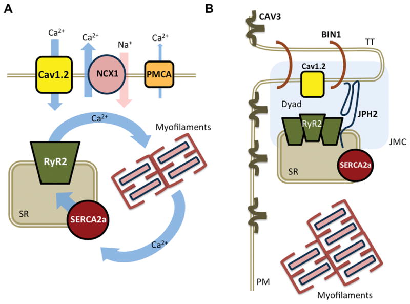
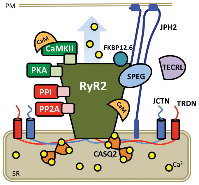
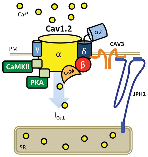
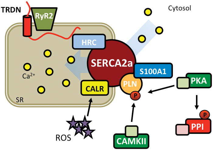
There has been a significant progress in our understanding of the molecular mechanisms by which calcium (Ca2+) ions mediate various types of cardiac arrhythmias. A growing list of inherited gene defects can cause potentially lethal cardiac arrhythmia syndromes, including catecholaminergic polymorphic ventricular tachycardia, congenital long QT syndrome, and hypertrophic cardiomyopathy. In addition, acquired deficits of multiple Ca2+-handling proteins can contribute to the pathogenesis of arrhythmias in patients with various types of heart disease. In this review article, we will first review the key role of Ca2+ in normal cardiac function-in particular, excitation-contraction coupling and normal electric rhythms. The functional involvement of Ca2+ in distinct arrhythmia mechanisms will be discussed, followed by various inherited arrhythmia syndromes caused by mutations in Ca2+-handling proteins. Finally, we will discuss how changes in the expression of regulation of Ca2+ channels and transporters can cause acquired arrhythmias, and how these mechanisms might be targeted for therapeutic purposes.

Keywords: arrhythmias, cardiac; atrial fibrillation; calcium channels; cardiomyopathy; ryanodine receptor calcium release channel.


Figures

Figure 1
Figure 1. Role of calcium-handling in excitation-contraction (EC) coupling
A. Schematic overview of key Ca2+-handling proteins involved in EC coupling. B. Schematic diagram of Ca2+ the release unit and major components of the JCM. The transverse tubule (TT) and SR membranes approximate to form the dyad. BIN1, bridging integrator 1; Cav1.2, L-type Ca2+ channel; CAV3, caveolin-3; JMC, junctional membrane complex; JPH2, juncophilin-2; NCX1, Na+/Ca2+ exchanger; PM, plasma membrane; PMCA, plasmalemmal Ca2+-ATPase; RyR2, ryanodine receptor type-2; SERCA2a, sarco/endoplasmic reticulum ATPase type-2a; SR, sarcoplasmic reticulum.
Figure 2
Figure 2. RyR2 macromolecular complex
Cartoon representing RyR2 pore-forming subunits with accessory proteins that bind to and/or modulate channel function. CaM, calmodulin; CaMKII, Ca2+/calmodulin-dependent protein kinase II; CASQ2, calsequestrin-2; FKBP12.6, FK506-binding protein 12.6; JCTN, junctin; JPH2, juncophilin-2; PKA, protein kinase A; PM, plasma membrane; PP, protein phosphatase; SR, sarcoplasmic reticulum; TECRL, trans-2,3-enoyl-CoA reductase-like protein; TRDN; triadin.
Figure 3
Figure 3. LTCC macromolecular complex
Cartoon representing Cav1.2 pore-forming β subunit with accessory β2, β, γ, δ subunits. CaMKII, Ca2+/calmodulin-dependent protein kinase II; CAV3, caveolin-3; ICa,L, L-type Ca2+ current; JPH2, juncophilin-2; PKA, protein kinase A; PM, plasma membrane; PP, protein phosphatase; SR, sarcoplasmic reticulum.
Figure 4
Figure 4. SERCA2a macromolecular complex
Cartoon representing SERCA2a complex required for reuptake of Ca2+ from the cytosol to the SR. CALR, calreticulin; CaMKII, Ca2+/calmodulin-dependent protein kinase II; HRC, histidine-rich Ca2+ binding protein; PKA, protein kinase A; PP, protein phosphatase; ROS, reactive oxygen species; RyR2, ryanodine receptor type-2; SERCA2a, sarco/endoplasmic reticulum ATPase type-2a; SR, sarcoplasmic reticulum; TRDN, triadin
Figure 5
Figure 5. Key electrophysiological mechanisms leading to cardiac arrhythmias
Ectopic (triggered) activity is primarily caused by A) early after-depolarizations (EADs) that occur mainly during bradycardia or following a pause, and B) delayed after-depolarizations (DADs) that occur using during tachycardia. Reentry requires a vulnerable substrate, which can be caused by C) action potential shortening or D) dispersion of refractoriness. ICa,L, L-type Ca2+ current; IK,Ca, Ca2+ dependent K+ current; INa,L, late Na+ current; INCX, Na+/Ca2+ exchanger current;
Figure 6
Figure 6. Diagram showing which genes have been linked to genetic arrhythmia disorders
Yellow fill indicates gene that encodes a Ca2+-sensitive or Ca2+-handling protein. CPVT, catecholaminergic polymorphic ventricular tachycardia; IVF, idiopathic ventricular fibrillation.
Figure 7
Figure 7. Calcium-dependent arrhythmia mechanisms in atrial fibrillation (AF)
Schematic diagram delineating which changes in intracellular Ca2+-handling promote arrhythmia mechanisms leading to AF. Enhanced RyR2-mediated Ca2+ release leads to activation of NCX, which in turn can cause a DAD-mediated triggered action potential (AP). Shortening of the AP duration (APD) due to reduction of ICa,L (L-type Ca2+ current) and membrane hyperpolarization due to upregulation of IK,1 (inward rectifier K+ current) promote reentry. CaM, calmodulin; CaMKII, Ca2+/calmodulin-dependent protein kinase II; Cav1.2, L-type Ca2+ channels; Cn, calcineurin; FKBP12.6, FK506-binding protein 12.6; miR-26, micro-RNA-26; mRNA, messenger RNA; NFAT, nuclear factor of activated T-cells; RyR2, ryanodine receptor type-2; SERCA2a, sarco/endoplasmic reticulum ATPase type-2a.

Similar articles

Cited by

References

    1. Cheng H, Lederer WJ. Calcium sparks. Physiol Rev. 2008;88:1491–1545. - PubMed
    1. Ringer S. A further Contribution regarding the influence of the different Constituents of the Blood on the Contraction of the Heart. J Physiol. 1883;4:29–42. - PMC - PubMed
    1. Bers DM. Cardiac excitation-contraction coupling. Nature. 2002;415:198–205. - PubMed
    1. Heijman J, Voigt N, Wehrens XH, Dobrev D. Calcium dysregulation in atrial fibrillation: the role of CaMKII. Front Pharmacol. 2014;5:30. - PMC - PubMed
    1. Bers DM. Excitation-Contraction Coupling and Cardiac Contractile Force. Boston: Kluwer Academic Publisher; 2001.

MeSH terms

Substances