. 2017 Aug 1;130(15):2491-2505.
doi: 10.1242/jcs.201897. Epub 2017 Jun 8.

Disruption of Rab8a and Rab11a causes formation of basolateral microvilli in neonatal enteropathy

Affiliations

Disruption of Rab8a and Rab11a causes formation of basolateral microvilli in neonatal enteropathy

Qiang Feng et al. J Cell Sci. .

Erratum in

Abstract

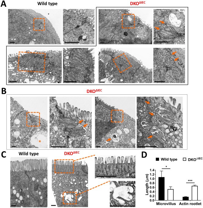
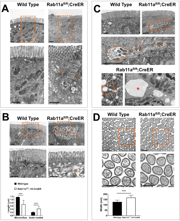
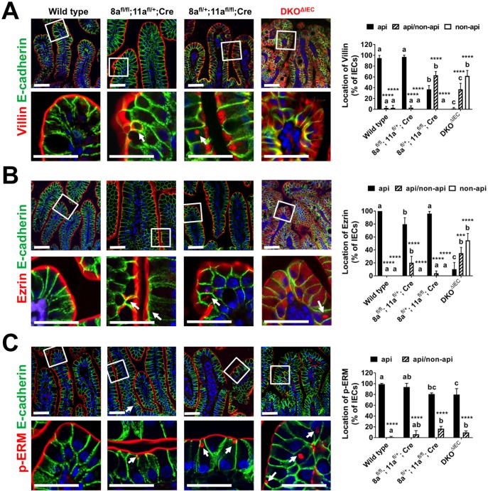
Misplaced formation of microvilli to basolateral domains and intracellular inclusions in enterocytes are pathognomonic features in congenital enteropathy associated with mutation of the apical plasma membrane receptor syntaxin 3 (STX3). Although the demonstrated binding of Myo5b to the Rab8a and Rab11a small GTPases in vitro implicates cytoskeleton-dependent membrane sorting, the mechanisms underlying the microvillar location defect remain unclear. By selective or combinatory disruption of Rab8a and Rab11a membrane traffic in vivo, we demonstrate that transport of distinct cargo to the apical brush border rely on either individual or both Rab regulators, whereas certain basolateral cargos are redundantly transported by both factors. Enterocyte-specific Rab8a and Rab11a double-knockout mouse neonates showed immediate postnatal lethality and more severe enteropathy than single knockouts, with extensive formation of microvilli along basolateral surfaces. Notably, following an inducible Rab11a deletion from neonatal enterocytes, basolateral microvilli were induced within 3 days. These data identify a potentially important and distinct mechanism for a characteristic microvillus defect exhibited by enterocytes of patients with neonatal enteropathy.

Keywords: Enterocyte; Microvillus formation; Neonatal enteropathy; Rab11a; Rab8a.


Conflict of interest statement

Competing interestsThe authors declare no competing or financial interests.

Figures

Fig. 1.
Fig. 1.
Selected or combined disruption of Rab8a and Rab11a traffic in IECs causes neonatal enteropathy of variable severities and microvillus hypotrophy. (A) Mice with IEC-specific deletion of both Rab8a and Rab11a (DKOΔIEC) displayed immediate postnatal lethality, demonstrating a more severe disease manifestation than mice that retained a single Rab8a or Rab11a allele in IECs. (B) 1-day-old DKOΔIEC and Rab8afl/+;Rab11afl/fl;Villin-Cre pups showed significantly lower body weight. Data are mean±s.e.m.; bars marked by the same letter (a or b) were not significantly different by one-way ANOVA. (C) DKOΔIEC neonates were smaller than wild-type littermates and had wrinkled skin, reflective of dehydration. Scale bar: 0.5 inches. (D) H&E staining of P1 duodenums, illustrating various degrees of villus hypotrophy in mutants. Scale bars: 200 μm. (E) PAS staining showed various degrees of presentation of PAS-positive intracellular vesicles (short arrow) in mutant IECs. Long arrows indicate fused villi. Scale bars: 50 μm. (F,G) Duodenums from 1-day-old wild-type and DKOΔIEC mice were analyzed by SEM. (H-K) SEM micrographs illustrating the more extensive microvillus deficiency in DKOΔIEC enterocytes residing in the upper villus, compared to those in the lower villus.
Fig. 2.
Fig. 2.
DKOΔIEC neonatal duodenum develops extensive lateral microvilli. (A) Duodenums from 1-day-old wild-type and DKOΔIEC mice were analyzed by TEM. DKOΔIEC enterocytes displayed intracellular accumulation of vesicles and formation of subapical microvilli (arrows) along the lateral sides of cells. Boxed areas are shown at higher magnification in insets. (B) Additional TEM micrographs of DKOΔIEC duodenal cells demonstrate the formation of lateral microvilli (arrows) underneath the tight junctions. (C) Representative TEM micrographs showing that some DKOΔIEC duodenal enterocytes had shorter apical microvilli, elongated microvillar actin rootlets, expanded terminal web, and presentation of MVID-typical vesicular body, with 1–2 microvillus projections facing the lumen. Scale bars: 1 μm. (D) Measurements of the lengths of microvilli and microvillar rootlets showed significant alterations in these structures in DKOΔIEC cells. Data represent 20 enterocytes of each genotype from three independent animals. *P<0.05, ***P<0.001.
Fig. 3.
Fig. 3.
Inducible deletion of Rab11a from neonatal IECs induces abnormal formation of lateral microvilli. (A) 3-day-old wild-type and Rab11afl/fl;Villin-CreER pups were administrated one dose of tamoxifen injection. Duodenums were analyzed by TEM 3 days after injection (n=2 for each genotype). Rab11afl/fl;Villin-CreER enterocytes displayed formation of lateral microvilli (arrows) underneath an expanded terminal web zone. (B) Rab11afl/fl;Villin-CreER enterocytes also exhibited accumulations of subapical vesicles (arrows), significantly shortened microvilli, and expanded terminal web. (C) Additional micrographs illustrating the scattered growth of microvilli along the lateral membrane, along with abnormal formation of large extracellular vacuoles (asterisk) in cross section. Scale bar: 1 μm. (D) Inducible deletion of Rab11a significantly increased the microvillar width in Rab11afl/fl;Villin-CreER cells. Scale bar: 200 nm. ****P<0.0001.
Fig. 4.
Fig. 4.
Selective or combined loss of Rab8a and Rab11a differentially impacts microvillus structural components. (A) Duodenal sections of 1-day-old mice of various genotypes were analyzed by confocal immunofluorescence for villin. Note that the strict apical villin localization was preferentially more dependent on Rab11a than Rab8a. However, loss of both Rab11a and Rab8a genes drastically dispersed its localization to basolateral or cytosolic regions. Cells with exclusive apical villin localization (api, black bars), with apical and non-apical villin localizations (api/non-api, striped bars), or with a total loss of apical localization (non-api, white bars) were scored and presented as a percentage, from ∼100 enterocytes in three different villi. Data represent 2–4 animals of each genotype. Significant differences between cellular compartments within the same genotype are indicated (****P<0.0001). When comparing cellular localizations across genotypes, bars marked by different letters (a, b or c) were significantly different in ANOVA analysis (P<0.05). (B) Ezrin appeared to be redundantly transported by Rab8a and Rab11a. Note that total loss of both Rab8a and Rab11a genes significantly dispersed ezrin from the apical domain. (C) Apical p-ERM localization was slightly but significantly weakened by Rab11a loss but not by Rab8a loss. Total loss of both Rab8a and Rab11a genes weakened but did not abolish p-ERM apical localizations. Note that non-apical inclusions of p-ERM were detected in all mutants (arrows). Arrows indicate non-apical inclusions. Scale bars: 50 μm.
Fig. 5.
Fig. 5.
Selective or combined loss of Rab8a and Rab11a differentially impacts apical Mst4, syndapin 2 and PKCζ localization. (A) Duodenal sections of 1-day-old mice of various genotypes were analyzed for Mst4 by confocal immunofluorescence. Note that the apical Mst4 localization was modestly affected by Rab11a loss but not by Rab8a loss. However, loss of both Rab8a and Rab11a genes dispersed its apical localization to cytosolic regions. Cells with apical and non-apical Mst4 localizations (striped bars), and cells with only non-apical localization (white bars), are presented as a percentage from ∼100 enterocytes in three different villi. Data represent 2–4 different animals of each genotype. (B) Similar to Mst4, apical syndapin 2 localization was modestly affected by Rab11a loss and severely affected by total loss of both Rab8a and Rab11a genes. (C) Apical PKCζ localization was preferentially weakened by Rab8a loss but not by Rab11a loss. Scale bars: 50 μm. *P<0.05, ****P<0.0001.
Fig. 6.
Fig. 6.
Brush border localizations of AP, SI and NHE3 are sensitive to loss of either Rab8a or Rab11a traffic. (A) Duodenal sections of 1-day-old mice of various genotypes were analyzed for AP by confocal immunofluorescence. Note that the strict apical AP localization was affected by loss of either Rab8a or Rab11a. However, loss of both Rab8a and Rab11a genes most severely dispersed its apical localization to cytosolic regions. (B) Post-weaning duodenal sections of mice of various genotypes were analyzed for SI by confocal immunofluorescence. Similar to AP, SI brush border localization was disturbed by loss of either Rab8a or Rab11a. (C) Apical NHE3 localization showed similar patterns of change to AP and SI. Scale bars: 50 μm. *P<0.05, **P<0.01, ***P<0.001 and ****P<0.0001.
Fig. 7.
Fig. 7.
Rab8a and Rab11a redundantly traffic Na+/K+-ATPases and integrin β1 to basolateral domains. (A) Co-immunostaining for laminin and E-cadherin showed overall normal localization of E-cadherin. Note that there were occasional colocalizations between two proteins in DKOΔIEC tissues (arrows). (B) Na+/K+-ATPases were localized to basolateral domains in single knockouts, but became slightly diffuse in DKOΔIEC tissues. (C) Integrin β1 basolateral localization was modestly affected by Rab11a loss but not by Rab8a loss. However, total loss of both Rab8a and Rab11a genes dispersed its basolateral localization. (D) Western blot analysis of relevant apical and basolateral proteins using P1 duodenum tissue lysates of W and DKOΔIEC mice. Results represent at least two independent experiments for each protein marker. Scale bars: 30 μm. (E) Schematic illustrating the observed trafficking and microvillus defects in single- and double-knockout IECs. *P<0.05; **P<0.01; ***P<0.001; ****P<0.0001.

Similar articles

Cited by

References

    1. Achler C., Filmer D., Merte C. and Drenckhahn D. (1989). Role of microtubules in polarized delivery of apical membrane proteins to the brush border of the intestinal epithelium. J. Cell Biol. 109, 179-189. 10.1083/jcb.109.1.179 - DOI - PMC - PubMed
    1. Ameen N. A. and Salas P. J. I. (2000). Microvillus inclusion disease: a genetic defect affecting apical membrane protein traffic in intestinal epithelium. Traffic 1, 76-83. 10.1034/j.1600-0854.2000.010111.x - DOI - PubMed
    1. Ang A. L., Fölsch H., Koivisto U.-M., Pypaert M. and Mellman I. (2003). The Rab8 GTPase selectively regulates AP-1B-dependent basolateral transport in polarized Madin-Darby canine kidney cells. J. Cell Biol. 163, 339-350. 10.1083/jcb.200307046 - DOI - PMC - PubMed
    1. Apodaca G., Gallo L. I. and Bryant D. M. (2012). Role of membrane traffic in the generation of epithelial cell asymmetry. Nat. Cell Biol. 14, 1235-1243. 10.1038/ncb2635 - DOI - PMC - PubMed
    1. Aumailley M., Pesch M., Tunggal L., Gaill F. and Fassler R. (2000). Altered synthesis of laminin 1 and absence of basement membrane component deposition in (beta)1 integrin-deficient embryoid bodies. J. Cell Sci. 113, 259-268. - PubMed

MeSH terms