. 2017 Sep 1;123(3):544-557.
doi: 10.1152/japplphysiol.00242.2017. Epub 2017 Jun 8.

Pretreatment with indomethacin results in increased heat stroke severity during recovery in a rodent model of heat stroke

Affiliations

Pretreatment with indomethacin results in increased heat stroke severity during recovery in a rodent model of heat stroke

Gerald N Audet et al. J Appl Physiol (1985). .

Abstract

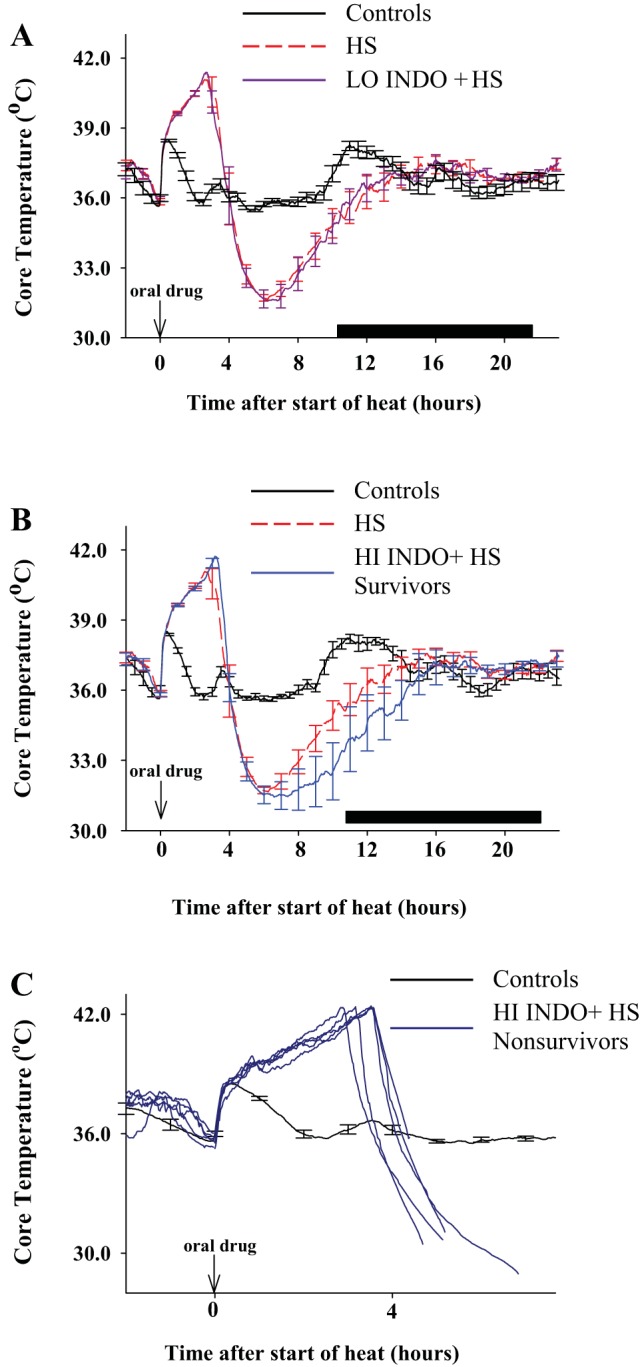
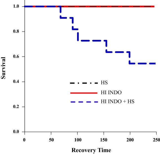
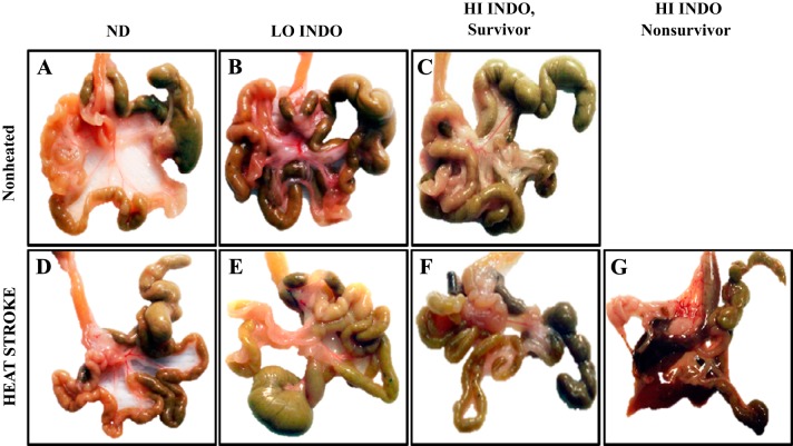
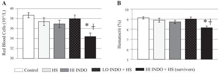
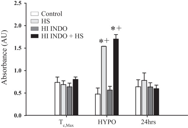
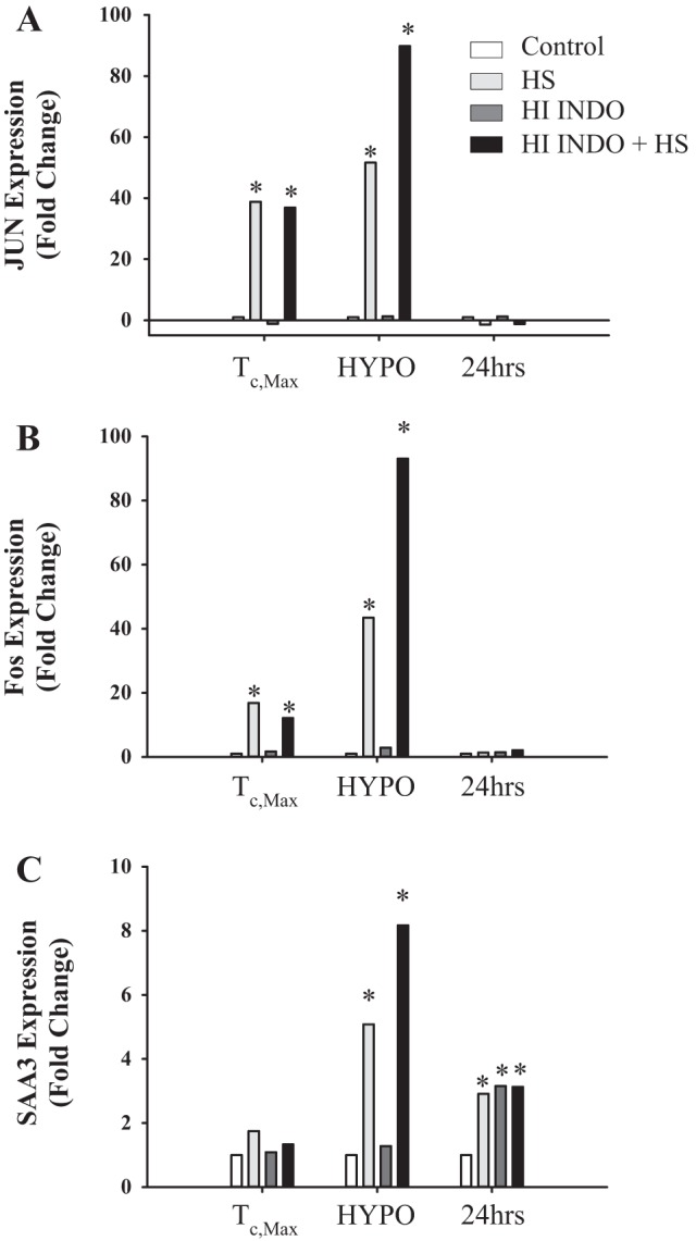
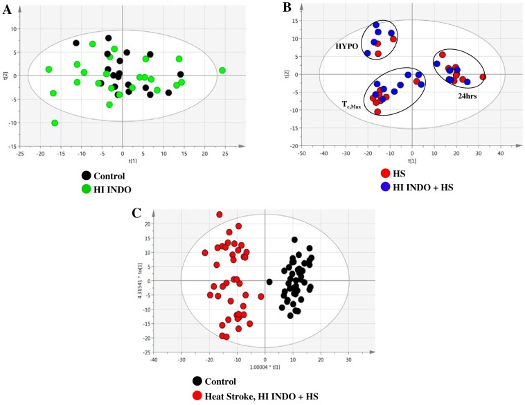
It has been suggested that medications can increase heat stroke (HS) susceptibility/severity. We investigated whether the nonsteroidal anti-inflammatory drug (NSAID) indomethacin (INDO) increases HS severity in a rodent model. Core temperature (Tc) of male, C57BL/6J mice (n = 45) was monitored continuously, and mice were given a dose of INDO [low dose (LO) 1 mg/kg or high dose (HI) 5 mg/kg in flavored treat] or vehicle (flavored treat) before heating. HS animals were heated to 42.4°C and euthanized at three time points for histological, molecular, and metabolic analysis: onset of HS [maximal core temperature (Tc,Max)], 3 h of recovery [minimal core temperature or hypothermia depth (HYPO)], and 24 h of recovery (24 h). Nonheated (control) animals underwent identical treatment in the absence of heat. INDO (LO or HI) had no effect on physiological indicators of performance (e.g., time to Tc,Max, thermal area, or cooling time) during heating or recovery. HI INDO resulted in 45% mortality rate by 24 h (HI INDO + HS group). The gut showed dramatic increases in gross morphological hemorrhage in HI INDO + HS in both survivors and nonsurvivors. HI INDO + HS survivors had significantly lower red blood cell counts and hematocrit suggesting significant hemorrhage. In the liver, HS induced cell death at HYPO and increased inflammation at Tc,Max, HYPO, and 24 h; however, there was additional effect with INDO + HS group. Furthermore, the metabolic profile of the liver was disturbed by heat, but there was no additive effect of INDO + HS. This suggests that there is an increase in morbidity risk with INDO + HS, likely resulting from significant gut injury.NEW & NOTEWORTHY This paper suggests that in a translational mouse model, NSAIDs may be counterindicated in situations that put an individual at risk of heat injury. We show here that a small, single dose of the NSAID indomethacin before heat stroke has a dramatic and highly damaging effect on the gut, which ultimately leads to increased systemic morbidity.

Keywords: NSAID; heat stress; heat stroke; hyperthermia; mouse.


Figures

Fig. 1.
Fig. 1.
Experimental outline. As described previously (63), animals were implanted with a small, wireless radiotelemetry device to monitor Tc responses >7 days before heating. The animals were placed in a room temperature (RT; 25 ± 2°C) incubator for 16–20 h before heating to acclimatize to incubator environment. At approximately 0700–1000, animals were weighed and given the drug or vehicle via a small bacon-flavored treat. Full consumption of the treat was confirmed while the animals were placed in incubator in the absence of normal food or water and the Ta increased to 39.5 ± 0.2°C. Once the animals reached a Tc,Max of 42.4°C, they were removed from the incubator and either euthanized at Tc,Max (I) or returned to RT for recovery. A subset of animals were then euthanized at hypothermic depth (~3 h; II) or 24 h post-Tc,Max (III). Nonheated animals (control, INDO) were subjected to the same time course as heated animals, except they remained at RT. The vehicle is a small bacon-flavored treat without INDO.
Fig. 2.
Fig. 2.
Tc profiles suggest no alterations in heating performance with HI INDO + HS, but significant detrimental alterations during recovery. Previously (63), mice were shown to have a biphasic Tc response during recovery from HS. After the rise in Tc associated with heating, there is a rapid cooling followed by a prolonged period of hypothermia. This is then followed by a return to baseline and then a ~1°C hyperthermic response after ~24 h (not shown). A: with LO INDO, there was no alteration in heating or recovery Tc responses compared with HS alone. B: this same response can be seen in HI INDO + HS survivors; however, the period of hypothermia is protracted compared with HS alone or LO INDO + HS, indicative of greater HS severity. C: nonsurvivors show in-heat responses identical to those of survivors; however, they enter hypothermia and are unable to stabilize Tc and succumb to HS post-Tc,Max. This suggests that there is no performance enhancement or detriment with INDO treatment during heating; however, there seems to be an increase in severity during recovery with HI INDO + HS treatment. Dark bars represent “lights off,” which is strictly controlled on a 12:12-h light-to-dark ratio.
Fig. 3.
Fig. 3.
Kaplan-Meier curve showing that HI INDO + HS animals have higher mortality rates compared with HI INDO or HS alone. Despite having similar in-heat parameters, HI INDO + HS animals (n = 11) had a 45% increase in mortality compared with HS (n = 9) or HI INDO (n = 9). Animals began to succumb at ~1 h post-start-of-heat, and survival rate continued to decrease through 4 h. All animals that did survive past ~4 h (250 min) survived through 24 h unless killed for analysis at HYPO. There was no mortality in any other groups.
Fig. 4.
Fig. 4.
Representative photographs of gross gut morphology across all treatment groups at 24 h of recovery. LO INDO in absence of heat resulted in no apparent gut damage (AC). Heat alone or with LO INDO also appeared to do no gross morphological damage (D and E). In the HI INDO + HS survivors (F) there is visual evidence for severely dehydrated intestinal and cecum content; however, there appears to be no hemorrhage or significant morphological damage. In the HI INDO + HS nonsurvivors (G) there was dramatic hemorrhage and significant tissue disruption throughout all sections of the small intestine and cecum at time of death. This suggests that there is an additive pathological load on the gut caused by the interaction of HI INDO and HS.
Fig. 5.
Fig. 5.
A decrease in RBC count and hematocrit at 24 h suggests significant blood loss in HI INDO + HS survivors. There was no difference in HI INDO or control animals. A: at 24 h, HS (n = 8, light gray bars) and LO INDO + HS (n = 13, patterned dark gray bars) did not alter RBC content compared with controls. However, there was a significant decrease (11%) in the HI INDO + HS (n = 10, black bars) survivors compared with controls (n = 8, white bars). B: there was a similar response in hematocrit, where HS and LO INDO + HS did not alter hematocrit; however, HI INDO + HS survivors showed a lower hematocrit (4.3%) compared with controls and HI INDO combined. These data suggest bleeding during recovery in HI INDO + HS survivors. We were unable to perform this analysis on HI INDO + HS nonsurvivors because of lack of sample. Two-way ANOVA; *P ≤ 0.05 compared with controls, +P < 0.05 compared with HI INDO.
Fig. 6.
Fig. 6.
HS increases cell death at HYPO, however, there is a lack of additive effect of HI INDO + HS. There was no effect of HI INDO alone (n = 6–9) on cell death at any time point. However, HS (n = 3–9) and HI INDO + HS (n = 5–9) groups showed increased cell death at HYPO compared with both controls (n = 3–9) and HI INDO (n = 6–9). There was no difference in HS vs. HI INDO + HS across any time points. Two-way ANOVA; *P < 0.05 compared with controls, +P < 0.05 compared with HI INDO.
Fig. 7.
Fig. 7.
Jun, Fos, and SAA3 gene expression is disrupted by HS but generally unaffected by INDO treatment. A: Jun fold gene expression is increased in HS (n = 3–8) and HI INDO + HS (n = 5–9) compared with controls (n = 3–9) at both Tc,Max (HS 38.84, HI INDO + HS 36.90) and HYPO (HS 51.16, HI INDO + HS 89.86). There were no changes at 24 h. HI INDO alone (n = 6–9) had no effect across all time points. B: Fos fold gene expression is increased in HS and HI INDO + HS compared with controls at both Tc,Max (HI 16.84, HI INDO + HS 12.12) and HYPO (HI 43.41, HI INDO + HS 93.06). There were no changes at 24 h. HI INDO alone had no effect across all time points. C: SAA3 fold gene expression was increased in HS and HI INDO groups at both HYPO (HS 5.08, HI INDO + HS 8.17) and 24 h (HS 2.91, HI INDO + HS 3.13) compared with controls. SAA3 was also increased at 24 h in the HI INDO group (3.15) compared with controls. Two-way ANOVA; *P < 0.05 compared with controls.
Fig. 8.
Fig. 8.
Multivariate analyses for impact of INDO treatment on metabolic liver profiles. Unsupervised multivariate analysis (PCA) of liver samples from mice dosed with NSAID (HI INDO) and no drug (control) in the absence or presence of heating. A: model statistics for HI INDO or control mice for heat exposure are R2X(cum) = 0.966 and Q2(cum) = 0.824; cum, cumulative. B: PCA shows separation by time between Tc,Max, HYPO, and 24 h. Model statistics for HS-exposed mice are R2X(cum) = 0.897 and Q2(cum) = 0.857. C: pairwise supervised analysis (OPLS-DA) of nonheated (control and HI INDO) compared with HS-exposed mice (HS and HI INDO + HS) demonstrating clear differentiation between the groups based on heat exposure. Model statistics are R2X(cum) = 0.946, R2Y(cum) = 0.907, and Q2(cum) = 0.805. Control, n = 4–8; HI INDO, n = 6–9; HS, n = 4–9; HI INDO + HS, n = 5–9.

Similar articles

Cited by

References

    1. Abate C, Patel L, Rauscher FJ III, Curran T. Redox regulation of fos and jun DNA-binding activity in vitro. Science 249: 1157–1161, 1990. doi:10.1126/science.2118682. - DOI - PubMed
    1. Abramson SB, Weissmann G. The mechanisms of action of nonsteroidal antiinflammatory drugs. Arthritis Rheum 32: 1–9, 1989. doi:10.1002/anr.1780320102. - DOI - PubMed
    1. Alván G, Orme M, Bertilsson L, Ekstrand R, Palmér L. Pharmacokinetics of indomethacin. Clin Pharmacol Ther 18: 364–373, 1975. doi:10.1002/cpt1975183364. - DOI - PubMed
    1. Audet GN, Dineen SM, Quinn CM, Leon LR. Altered hypothalamic inflammatory gene expression correlates with heat stroke severity in a conscious rodent model. Brain Res 1637: 81–90, 2016. doi:10.1016/j.brainres.2016.01.048. - DOI - PubMed
    1. Audet GN, Quinn CM, Leon LR. Point-of-care cardiac troponin test accurately predicts heat stroke severity in rats. Am J Physiol Regul Integr Comp Physiol 309: R1264–R1272, 2015. doi:10.1152/ajpregu.00286.2015. - DOI - PubMed

MeSH terms

Substances

LinkOut - more resources