. 2017 Sep;102(9):1594-1604.
doi: 10.3324/haematol.2016.163337. Epub 2017 Jun 8.

Extracellular vesicles of bone marrow stromal cells rescue chronic lymphocytic leukemia B cells from apoptosis, enhance their migration and induce gene expression modifications

Affiliations

Extracellular vesicles of bone marrow stromal cells rescue chronic lymphocytic leukemia B cells from apoptosis, enhance their migration and induce gene expression modifications

Emerence Crompot et al. Haematologica. 2017 Sep.

Abstract

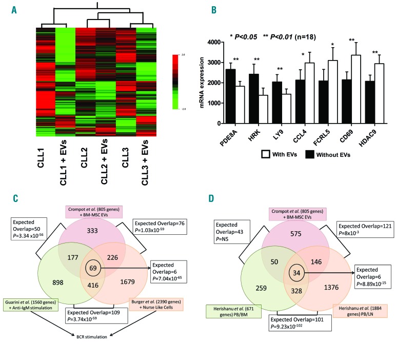
Interactions between chronic lymphocytic leukemia (CLL) B cells and the bone marrow (BM) microenvironment play a major function in the physiopathology of CLL. Extracellular vesicles (EVs), which are composed of exosomes and microparticles, play an important role in cell communication. However, little is known about their role in CLL / microenvironment interactions. In the present study, EVs purified by ultracentrifugation from BM mesenchymal stromal cell (BM-MSC) cultures were added to CLL B cells. After their integration into CLL B cells, we observed a decrease of leukemic cell spontaneous apoptosis and an increase in their chemoresistance to several drugs, including fludarabine, ibrutinib, idelalisib and venetoclax after 24 hours. Spontaneous (P=0.0078) and stromal cell-derived factor 1α -induced migration capacities of CLL B cells were also enhanced (P=0.0020). A microarray study highlighted 805 differentially expressed genes between leukemic cells cultured with or without EVs. Of these, genes involved in the B-cell receptor pathway such as CCL3/4, EGR1/2/3, and MYC were increased. Interestingly, this signature presents important overlaps with other microenvironment stimuli such as B-cell receptor stimulation, CLL/nurse-like cells co-culture or those provided by a lymph node microenvironment. Finally, we showed that EVs from MSCs of leukemic patients also rescue leukemic cells from spontaneous or drug-induced apoptosis. However, they induce a higher migration and also a stronger gene modification compared to EVs of healthy MSCs. In conclusion, we show that EVs play a crucial role in CLL B cells/BM microenvironment communication.


Figures

Figure 1.
Figure 1.
Characterization of bone marrow (BM) extracellular vesicles (EVs) and uptake by chronic lymphocytic leukemia (CLL) B cells. Bone marrow mesenchymal stromal cells (BM-MSCs) were characterized by electron microscopy (A) and flow cytometry (B) using the latex bead technique. EVs showed a classical spherical appearance by transmission electron microscopy (TEM). EVs expressed CD63 (EV marker) and CD73 (MSC marker) and were negative for CD45 (hematopoietic marker). (C) CLL B cells were incubated with BM-MSC EVs, previously labeled with PKH67, for 10 minutes (min), 1 hour (h), 3 h and 24 h. Flow cytometry showed a rapid increase of the mean of fluorescence intensity (MFI), depending on the incubation time. The uptake of BM-MSC EVs was a fast process; after 10 min, more than 60% of CLL B cells had integrated fluorescent vesicles. PBS: phosphate-buffered saline.
Figure 2.
Figure 2.
Bone marrow (BM) extracellular vesicles (EVs) protect chronic lymphocytic leukemia (CLL) B cells from spontaneous apoptosis. CLL B cells were incubated with bone marrow mesenchymal stromal cell (BM-MSC) EVs, and after 24 hours (h), cells were stained with annexin V-FITC/7AAD. We observed a decrease in spontaneous apoptosis (A) with a significant effect on early (B) and late (C) apoptosis. (D) A representative flow cytometry analysis of CLL B cells cultured with EVs for 24 h. (E) Expression of the anti-apoptotic protein Mcl-1 (myeloid cell leukemia 1). Red line: CLL B cells without EVs; blue line: CLL B cells cultured with EVs. The expression of secondary antibody was used as a negative control (n=10, P=0.0006). Results were also confirmed by qPCR (n=25, P=0.0001) (F).
Figure 3.
Figure 3.
Bone marrow (BM) extracellular vesicles (EVs) increase chronic lymphocytic leukemia (CLL) B-cell migration capacity. CLL B cells were incubated with bone marrow mesenchymal stromal cell (BM-MSC) EVs in a Transwell assay. EV addition increased the spontaneous migration (A). The migration index was also calculated as the number of cells transmigrating in the presence of SDF-1α divided by the number of cells transmigrating in the absence of SDF1α. A significant increase in migration was observed in the presence of the chemoattractant SDF-1α (B) (n=10, P=0.0020). (C) Migration of CLL B cells in the presence of AMD3100 and pertussis toxin (P. tox). We did not observe a decrease in the migrating cells in the presence of AMD3100 or pertussis toxin (P. tox).
Figure 4.
Figure 4.
Bone marrow (BM) extracellular vesicles (EVs) increase chemoresistance of chronic lymphocytic leukemia (CLL) B cells. CLL B cells were incubated with bone marrow mesenchymal stromal cell (BM-MSC) EVs for 4 hours (h) and then treated with drugs for 24 h: bortezomib (A), cladribine (B), fludarabine (C), flavopiridol (D), methylprednisolone (E), ibrutinib (F), idelalisib (G) and venetoclax (H). Cells were then stained with annexin V-FITC/7AAD. The addition of BM-MSC EVs in CLL B-cell culture could protect them from drug-induced apoptosis; we observed a decrease in annexin-positive cells in the presence of each of the 8 drugs. The results were normalized by the subtraction of spontaneous apoptosis (cells without drug and EVs).
Figure 5.
Figure 5.
Impact of bone marrow (BM) extracellular vesicles (EVs) on chronic lymphocytic leukemia (CLL) B-cell gene expression profile and comparison with other microarray studies. The genetic profiles of CLL B cells from 3 different patients were determined to obtain a global view of gene expression differences in leukemic cells with or without bone marrow mesenchymal stromal cell (BM-MSC) EV treatment. (A) A total of 805 genes were differentially expressed between leukemic cells cultured with or without EVs (P<0.05). (B) We selected 7 differentially expressed genes in our microarray analysis and confirmed their expression by real-time PCR. All genes were validated, confirming our microarray results. (C) We compared our microarray results with 2 other published studies on CLL B cells activated by NLC culture or IgM stimulation. We obtained a substantial overlap between the differentially expressed genes; 177 and 226 genes were shared between our study and those of Burger et al. and Guarini et al., respectively, and 69 genes were common among all 3 studies. (D) Our microarray results were also compared with the study of Herishanu et al. who analyzed the differential gene expression of CLL cells obtained from different compartments: peripheral blood (PB), bone marrow (BM) and lymph node (LN). A significant overlap was observed between an EV treatment and a stimulation provided by an LN microenvironment. In addition, 34 genes were in common between our study and CLL cells receiving stimuli from BM and LN (P=8.89×10⁻¹⁵). The “SuperExactTest” was applied to evaluate the statistical value of the intersections between 3 studies as indicated by the expected overlap and the P values. BCR: B-cell receptor.
Figure 6.
Figure 6.
Extracellular vesicles (EV) effects on chronic lymphocytic leukemia (CLL) B-cell receptor (BCR) activation. (A) Calcium flux in Raji cell line (n=3) after anti-IgM or EV treatment measured using FLUO8 by flow cytometry. The mean of fluorescence intensity (MFI) was normalized by the MFI of untreated cells and plotted every ten seconds. The phosphorylation of ERK (B and D) and AKT (C and E) was evaluated by flow cytometry. Data were presented as the mean fluorescence intensity (MFI) ratio of IgM stimulation/unstimulated cells and EVs treatment/untreated cells. Downstream targets of BCR activation in CLL B cells after EV integration were analyzed by real-time PCR (MYC, LPL, CCL4) (F–H) and flow cytometry (CD69, CXCR4 or CD184) (G). (H) Four targets of BCR activation (CCL4, miR-229c, miR-150, miR21) after 4 hours (h) or 24 h of EV integration treated or not with actinomycin D (5 μg/mL). Genes were normalized using cyclophilin A (PPIA) as endogenous control while microRNAs were normalized using RNU48. (I) CLL B cells were incubated with BM-MSC EVs in a Transwell assay. Cells were treated with or without ibrutinib (ibrut.: 10 μM) or idelalisib (idelal.: 10 μM), with or without EVs. The migration ratio represents the number of migrating cells in each condition divided by the spontaneous migrating cells (in absence of EVs or drugs). (J) Cells were treated with or without ibrutinib (10 μM) and with or without EVs. Gene expression for a selection of genes was evaluated by real-time PCR. Data were normalized with the expression of untreated cells (without EVs and without ibrutinib).
Figure 7.
Figure 7.
Quantitative and qualitative comparison between healthy and chronic lymphocytic leukemia (CLL)-derived extracellular vesicles (EVs). (A) Measure of mean size of EVs from healthy and CLL MSC. (B and C) A representative size distribution of healthy and CLL mesenchymal stromal cell (MSCs), respectively. CLL B cells were incubated with healthy or CLL-MSC EVs for 24 hours (h) and cells were stained with annexin V-FITC/7AAD to measure apoptosis (E) or DioC6/propidium iodide to measure cell viability (F). We observed a decrease of spontaneous apoptosis. CLL B cells were incubated with healthy or CLL-MSC EVs for 4 h and then treated for 24 h with ibrutinib (G), idelalisib (H) or venetoclax (I). Cells were then stained with annexin V-FITC/7AAD. The results were normalized by the subtraction of spontaneous apoptosis (cells without drug and EVs). (J) CLL B cells were incubated with healthy or CLL MSC EVs in a Transwell assay. The migration ratio represents the number of migrating cells in each condition divided by the spontaneous migrating cells (in absence of EVs). (K) Cells were incubated with healthy or CLL MSC EVs for 24 h. Gene expression for a selection of genes was evaluated by real-time PCR. Data were normalized with the expression of untreated cells (without EVs).

Similar articles

Cited by

References

    1. Ten Hacken E, Burger JA. Microenvironment dependency in Chronic Lymphocytic Leukemia: The basis for new targeted therapies. Pharmacol Ther. 2014;144(3):338–348. - PubMed
    1. Lagneaux L, Delforge A, Bron D, De Bruyn C, Stryckmans P. Chronic lymphocytic leukemic B cells but not normal B cells are rescued from apoptosis by contact with normal bone marrow stromal cells. Blood. 1998;91(7):2387–2396. - PubMed
    1. Raposo G, Stoorvogel W. Extracellular vesicles: exosomes, microvesicles, and friends. J Cell Biol. 2013;200(4):373–383. - PMC - PubMed
    1. Lotvall J, Hill AF, Hochberg F, et al. Minimal experimental requirements for definition of extracellular vesicles and their functions: a position statement from the International Society for Extracellular Vesicles. J Extracell Vesicles. 2014;3:26913. - PMC - PubMed
    1. Abels ER, Breakefield XO. Introduction to Extracellular Vesicles: Biogenesis, RNA Cargo Selection, Content, Release, and Uptake. Cell Mol Neurobiol. 2016; 36(3):301–312. - PMC - PubMed

Publication types

MeSH terms