. 2017 Sep;102(9):1519-1529.
doi: 10.3324/haematol.2016.163436. Epub 2017 Jun 8.

Combined targeting of STAT3 and STAT5: a novel approach to overcome drug resistance in chronic myeloid leukemia

Affiliations

Combined targeting of STAT3 and STAT5: a novel approach to overcome drug resistance in chronic myeloid leukemia

Karoline V Gleixner et al. Haematologica. 2017 Sep.

Abstract

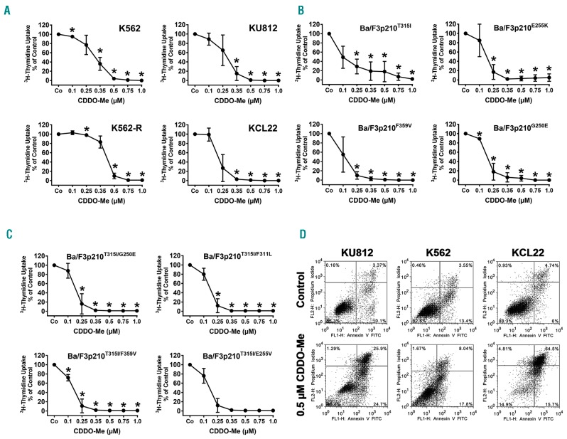
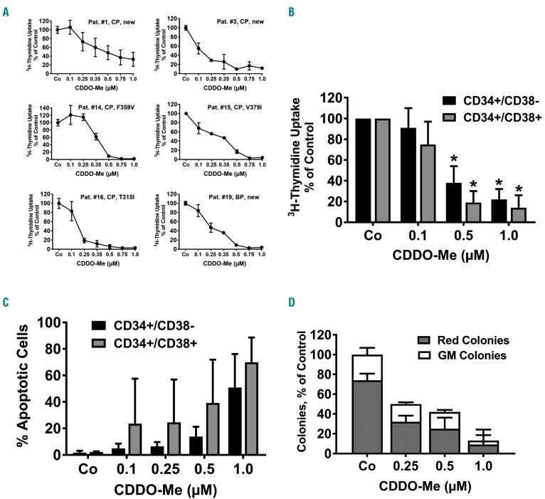
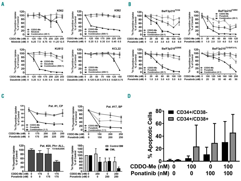
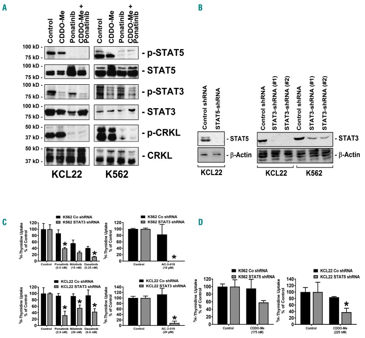
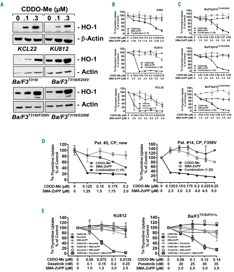
In chronic myeloid leukemia, resistance against BCR-ABL1 tyrosine kinase inhibitors can develop because of BCR-ABL1 mutations, activation of additional pro-oncogenic pathways, and stem cell resistance. Drug combinations covering a broad range of targets may overcome resistance. CDDO-Me (bardoxolone methyl) is a drug that inhibits the survival of leukemic cells by targeting different pro-survival molecules, including STAT3. We found that CDDO-Me inhibits proliferation and survival of tyrosine kinase inhibitor-resistant BCR-ABL1+ cell lines and primary leukemic cells, including cells harboring BCR-ABL1T315I or T315I+ compound mutations. Furthermore, CDDO-Me was found to block growth and survival of CD34+/CD38- leukemic stem cells (LSC). Moreover, CDDO-Me was found to produce synergistic growth-inhibitory effects when combined with BCR-ABL1 tyrosine kinase inhibitors. These drug-combinations were found to block multiple signaling cascades and molecules, including STAT3 and STAT5. Furthermore, combined targeting of STAT3 and STAT5 by shRNA and STAT5-targeting drugs also resulted in synergistic growth-inhibition, pointing to a new efficient concept of combinatorial STAT3 and STAT5 inhibition. However, CDDO-Me was also found to increase the expression of heme-oxygenase-1, a heat-shock-protein that triggers drug resistance and cell survival. We therefore combined CDDO-Me with the heme-oxygenase-1 inhibitor SMA-ZnPP, which also resulted in synergistic growth-inhibitory effects. Moreover, SMA-ZnPP was found to sensitize BCR-ABL1+ cells against the combination 'CDDO-Me+ tyrosine kinase inhibitor'. Together, combined targeting of STAT3, STAT5, and heme-oxygenase-1 overcomes resistance in BCR-ABL1+ cells, including stem cells and highly resistant sub-clones expressing BCR-ABL1T315I or T315I-compound mutations. Whether such drug-combinations are effective in tyrosine kinase inhibitor-resistant patients with chronic myeloid leukemia remains to be elucidated.


Figures

Figure 1.
Figure 1.
CDDO-Me inhibits growth and viability of Philadelphia-positive (Ph+) cell lines. (A–C) Human chronic myeloid leukemia (CML) cell lines (A) and BCR-ABL1-expressing Ba/F3 cells (B and C) were exposed to control medium (Co) or various concentrations of CDDO-Me for 48 hours (h). In case of K562-R, imatinib was removed prior to CDDO-Me exposure. Then, proliferation was measured by assessing 3H-thymidine uptake. Results are expressed in % of control and represent the mean±Standard Deviation (S.D.) of 3 independent experiments. (D) CML cell lines (K562, KU812, KCL22) were incubated in control medium or in CDDO-Me (0.5 μM) for 48 h. Thereafter, the percentage of apoptotic cells was determined by combined Annexin V/PI staining. The figure shows dot blots from one representative experiment. Almost identical results were obtained in two other experiments.
Figure 2.
Figure 2.
CDDO-Me counteracts the proliferation of primary chronic myeloid leukemia (CML) cells. (A) Primary CML cells were isolated from the peripheral blood (PB) of 6 patients [chronic phase (CP) n=5; primary blast phase (BP) n=1]. In 3 patients, cells were obtained at diagnosis (“new”). In all 3 TKI-resistant patients, BCR-ABL1 mutations were detected as indicated. Isolated cells were incubated in control medium (Co) or various concentrations of CDDO-Me as indicated at 37°C for 48 hours (h). Then, proliferation was measured by assessing 3H-thymidine incorporation. Results are expressed in % of control and represent the mean±Standard Deviation (S.D.) of triplicates. Patients’ numbers refer to Table 1. (B) Highly purified CD34+/CD38 stem cells (black bars) and CD34+/CD38+ precursor cells (gray bars) were sorted from peripheral blood (PB) leukocytes of 3 patients (#9, #11 and #17) and were kept in control medium (Co) or various concentrations of CDDO-Me as indicated at 37°C for 48 h. Then, proliferation was measured by assessing 3H-thymidine incorporation. Results are expressed as % of control and represent the mean±S.D. of 3 patients. *P<0.05 compared to control (Co). (C) Primary PB mononuclear cells (MNC) were isolated from 3 patients (#9, #11, #17) and kept in control medium (Co) or various concentrations of CDDO-Me at 37°C for 48 h. Thereafter, cells were subjected to flow cytometry to determine the % of apoptotic (Annexin V+) CD34+/CD38 (stem) cells (black bars) and CD34+/CD38+ (progenitor) cells (gray bars). Results represent the mean±S.D. of 3 patients. (D) PB MNC from 3 patients (#9, #10, #11) were cultured in methylcellulose with cytokines in the absence (Co) or presence of various concentrations of CDDO-Me as indicated for 14 days. Then, the numbers of granulocyte/macrophage (GM) colonies (white bars) and red cell-containing (burst-forming plus erythroid) colonies (gray bars) were counted under an inverted microscope. Results are expressed in % of control (100% = red colonies + GM colonies in the absence of CDDO-Me) and represent the mean±S.D. of 3 patients.
Figure 3.
Figure 3.
CDDO-Me synergizes with BCR-ABL1 TKI in producing growth-inhibition in Philadelphia-positive (Ph+) cells. (A and B) Human chronic myeloid leukemia (CML) cell lines (A) or Ba/F3 cells harboring various BCR-ABL1 mutants (B) were incubated in control medium (0) or in various concentrations of CDDO-Me (●–●), BCR-ABL1-targeting TKI as indicated (■–■), or combinations of drugs at a fixed ratio of drug-concentrations as indicated (▲−▲) for 48 hours (h). Thereafter, 3H-thymidine incorporation was measured. Results are expressed in % of control and represent the mean± Standard Deviation (S.D.) of triplicates. (C) Primary neoplastic cells isolated from patient #1, #11, #17 and #20 as well as normal bone marrow (BM) mononuclear cells (MNC) obtained from 3 donors were incubated in various concentrations of CDDO-Me or ponatinib (as single drugs or in combination) as indicated. Results in the upper panels and the lower left panel are expressed in % of control and represent the mean±S.D. of triplicates. Results in the lower right panel show the mean±S.D. of 3 normal donors (gray bars) or 3 CML patients (#1, #11 and #17) (black bars). (D) Primary peripheral blood (PB) MNC were isolated from 3 patients (#9, #11, #17) and kept in control medium (0) or various concentrations of CDDO-Me or ponatinib as indicated at 37°C for 48 h. Thereafter, cells were subjected to flow cytometry to determine the % of apoptotic (Annexin V+) CD34+/CD38 (stem) cells (black bars) and CD34+/CD38+ (progenitor) cells (gray bars). Results represent the mean±S.D. of 3 patients.
Figure 4.
Figure 4.
Combined inhibition of STAT3 and STAT5 with shRNA and targeted drugs results in synergistic growth inhibition in chronic myeloid leukemia (CML) cells. (A) KCL22 and K562 cells were incubated with CDDO-Me (1 μM), ponatinib (1 μM) or a combination of both drugs for 4 hours (h). Thereafter, cells were subjected to Western blot analysis using antibodies against p-STAT5, STAT5, p-STAT3, STAT3, p-CRKL, or CRKL, as indicated. (B–D) K562 and KCL22 cells were transfected with control shRNA, with an shRNA-construct directed against STAT5, or shRNA-constructs directed against STAT3 (#1 = #V3LHS_376016; #2 = #V3LHS_641818) as indicated. Protein knockdown was confirmed by western blotting using antibodies against STAT3 or STAT5. β-Actin served as loading control (B). (C) K562 cells (upper panels) and KCL22 cells (lower panels) were treated with control-shRNA (black bars) or with shRNA directed against STAT3 (construct #2, gray bars) and were then incubated with control medium (control), with BCR-ABL1 TKI (as indicated), or with the STAT5 inhibitor AC-3-019 for 48 h. Results are expressed in % of control and represent the mean±Standard Deviation (S.D.) of triplicates. *P<0.05. (D) K562 cells (left panel) and KCL22 cells (right panel) were treated with control-shRNA (black bars) or shRNA against STAT5 (gray bars) and were then incubated in control medium or in CDDO-Me for 48 h. Results are expressed in % of control and represent the mean±S.D. of triplicates. *P<0.05.
Figure 5.
Figure 5.
SMA-ZnPP sensitizes BCR-ABL1+ cells against CDDO-Me and against the combination “CDDO-Me+BCR-ABL1 TKI”. (A) KCL22, KU812 and BCR-ABL1+ Ba/F3 cells were incubated in control medium or in 0.1 or 0.3 μM CDDO-Me for 24 hours (h). Then, cells were subjected to Western blot analysis using antibodies against HO-1 or either β-Actin or Actin (loading control) as indicated. (B–D) Cell lines (B and C) and primary chronic myeloid leukemia (CML) cells (D) were incubated in control medium or in various concentrations of CDDO-Me (●–●), SMA-ZnPP (■–■), or a combination of both drugs (at fixed ratio of drug concentrations) (▲−▲) for 48 hours (h). Then, 3H-thymidine incorporation was measured. Results are expressed in % of control and represent the mean±Standard Deviation (S.D.) of triplicates. Patient numbers in (D) refer to Table 1. (E) KU812 and Ba/F3p210T315I/F311L cells were exposed to low doses of CDDO-Me (●–●), a BCR-ABL1 TKI (dasatinib or ponatinib) (■–■), and SMA-ZnPP (▲−▲), either as single agents (blue graphs), in 2-drug combinations (green graphs; CDDO-Me+TKI: ▼–▼; CDDO-Me+SMA-ZnPP: ♦–♦; TKI+SMA-ZnPP: ○–○), or in a 3-drug combination (red graph; □–□) for 48 h. Then, 3H-thymidine uptake was measured. Results are expressed in % of control and represent the mean±S.D. of triplicates.

Similar articles

Cited by

References

    1. Nowell PC, Hungerford DA. A minute chromosome in human granulocytic leukemia. Science. 1960;132:1497.
    1. Rowley JD. A new consistent chromosomal abnormality in chronic myelogenous leukaemia identified by quinacrine fluorescence and Giemsa staining. Nature. 1973;243(5405):290–293. - PubMed
    1. Bedi A, Zehnbauer BA, Barber JP, Sharkis SJ, Jones RJ. Inhibition of apoptosis by BCR-ABL in chronic myeloid leukemia. Blood. 1994;83(8):2038–2044. - PubMed
    1. Druker BJ, Talpaz M, Resta DJ, et al. Efficacy and safety of a specific inhibitor of the BCR-ABL tyrosine kinase in chronic myeloid leukemia. N Engl J Med. 2001;344(14):1031–1037. - PubMed
    1. Kantarjian H, Sawyers C, Hochhaus A, et al. Hematologic and cytogenetic responses to imatinib mesylate in chronic myelogenous leukemia. N Engl J Med. 2002;346(9):645–652. - PubMed

Publication types

MeSH terms

Substances