. 2017 Sep;27(9):1487-1500.
doi: 10.1101/gr.211649.116. Epub 2017 Jun 8.

Yeast genetic interaction screen of human genes associated with amyotrophic lateral sclerosis: identification of MAP2K5 kinase as a potential drug target

Affiliations

Yeast genetic interaction screen of human genes associated with amyotrophic lateral sclerosis: identification of MAP2K5 kinase as a potential drug target

Myungjin Jo et al. Genome Res. 2017 Sep.

Abstract

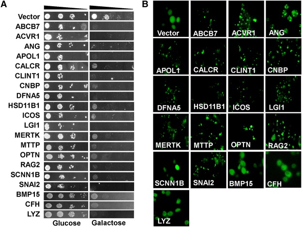
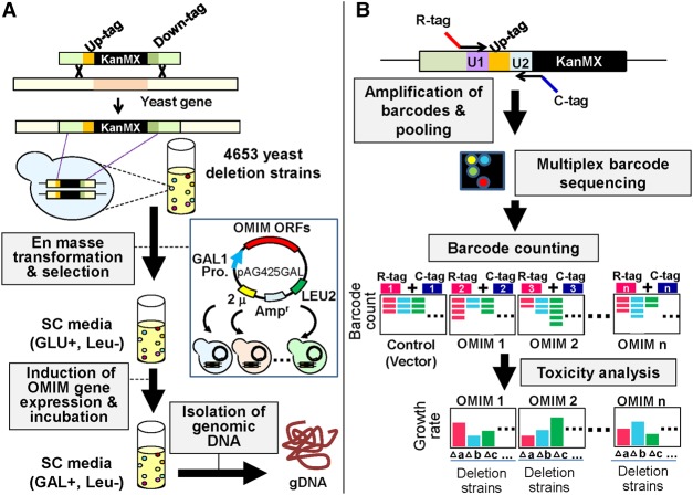
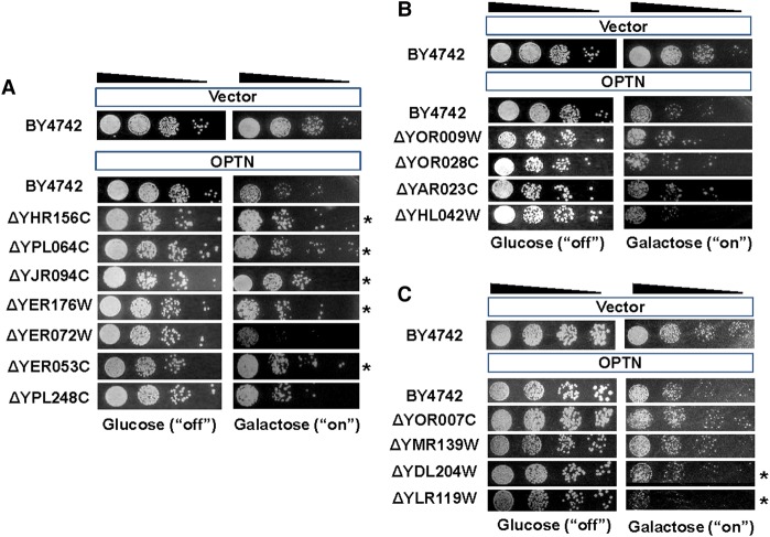
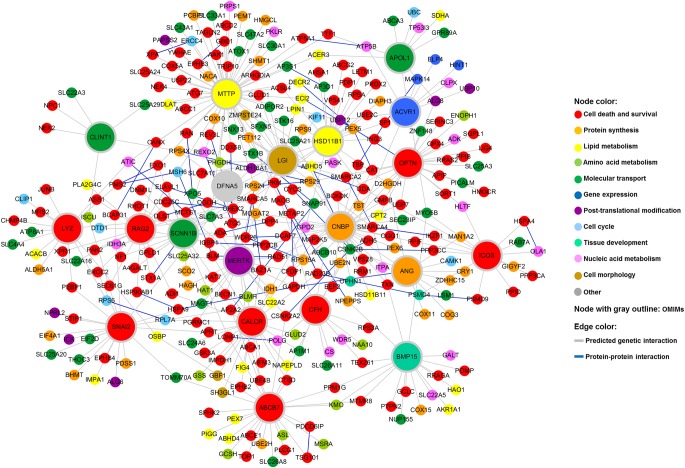
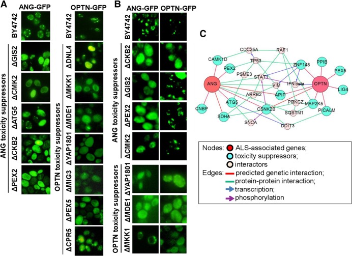
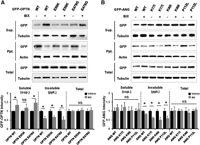
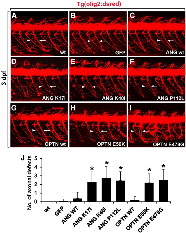
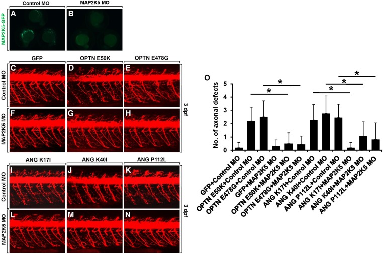
To understand disease mechanisms, a large-scale analysis of human-yeast genetic interactions was performed. Of 1305 human disease genes assayed, 20 genes exhibited strong toxicity in yeast. Human-yeast genetic interactions were identified by en masse transformation of the human disease genes into a pool of 4653 homozygous diploid yeast deletion mutants with unique barcode sequences, followed by multiplexed barcode sequencing to identify yeast toxicity modifiers. Subsequent network analyses focusing on amyotrophic lateral sclerosis (ALS)-associated genes, such as optineurin (OPTN) and angiogenin (ANG), showed that the human orthologs of the yeast toxicity modifiers of these ALS genes are enriched for several biological processes, such as cell death, lipid metabolism, and molecular transport. When yeast genetic interaction partners held in common between human OPTN and ANG were validated in mammalian cells and zebrafish, MAP2K5 kinase emerged as a potential drug target for ALS therapy. The toxicity modifiers identified in this study may deepen our understanding of the pathogenic mechanisms of ALS and other devastating diseases.


Figures

Figure 1.
Figure 1.
Overexpression of selected OMIM genes induces toxicity and cytoplasmic aggregates in yeast. (A) In total, 1305 OMIM ORFs were cloned under the control of a galactose-inducible promoter in pAG425 vectors. pAG425Gal-OMIMs were individually transformed into yeast. Transformants were grown in SRaf-Leu medium for 16 h, spotted onto SD-Leu agar plates (OMIM expression “off”) or SGal-Leu agar plates (OMIM expression “on”), and incubated for 3 d. Shown are 10-fold serial dilutions starting with an equal number of cells expressing the 20 toxic OMIM genes. Nontoxic OMIM genes are not shown. (B) Yeast cells expressing the “C-terminal GFP-tagged OMIM” fusion proteins were visualized with fluorescence microscopy. GFP alone was distributed in the cytoplasm. Most of the toxic GFP-tagged OMIM proteins formed protein aggregates.
Figure 2.
Figure 2.
Flowchart describing the yeast genetic interaction screen. (A) The 20 toxic OMIM genes were transformed individually into a pool of 4653 yeast homozygous deletion strains containing a 20-bp DNA barcode sequence. Transformants were selected in SD-Leu medium for 16 h and then washed twice with PBS. The cells were resuspended in SGal-Leu medium and incubated for 2 d to induce the expression of OMIM genes under the control of the GAL1 promoter. Genomic DNA was separately isolated from cells harvested at the end of pooled culture in the presence of GLU or GAL. (B) Barcodes were amplified from genomic DNA with multiplexed primers containing distinct combinations of two different tags for each OMIM gene. Equal amounts of DNA amplified for each OMIM gene were pooled and subjected to multiplex barcode sequencing using an Illumina Genome Analyzer. Next-generation sequencing data were then analyzed for barcode counting.
Figure 3.
Figure 3.
Validation of the genome-wide screening data for OPTN. Barcode counting was used to screen OMIM-yeast genetic interactions. From the Bar-seq data analysis and Z-score distributions, three groups of yeast genes were chosen for spotting assays: group 1, toxicity suppressors; group 2, no effects; and group 3, toxicity enhancers. OPTN toxicity was analyzed with spotting assays in the wild-type yeast strain (BY4742), toxicity-suppressing gene deletion strains (A), no-effect gene deletion strains (B), and toxicity-enhancing gene deletion strains (C). pAG425GAL-ccdB was used as the empty vector control. Twenty-four yeast deletions were tested. Representative results are shown. Asterisks indicate yeast deletions that suppressed or enhanced OPTN toxicity.
Figure 4.
Figure 4.
Human–yeast genetic interaction network. Human orthologs of yeast genes whose deletion suppressed the toxicity of the 20 OMIM ORFs were identified. A network view of these human orthologs was generated using Cytoscape. The node color corresponds to the biological function category to which the gene belongs. The color of an edge indicates the type of interaction.
Figure 5.
Figure 5.
Formation of ALS-associated protein aggregates is attenuated in specific yeast gene deletion strains. (A) Loss of specific yeast genes reversed the aggregation of the ALS-associated proteins. To observe protein localization, OPTN and ANG were cloned into pAG425GAL-ccdB-GFP and transformed into the deletion strains. Cells expressing OPTN-GFP or ANG-GFP were observed with fluorescence microscopy after 16 h of OPTN/ANG gene induction. (B) OPTN and ANG protein aggregation was cross-tested in representative yeast deletion strains of ANG and OPTN toxicity suppressors, respectively. (C) From the genetic interaction results of the cross-test, a network for the two ALS genes was constructed.
Figure 6.
Figure 6.
MAP2K5 inhibition attenuates the formation of OPTN and ANG protein aggregates in mammalian cells. NIH3T3 cells were transiently transfected with wild-type GFP-OPTN or GFP-OPTN mutants (E50K or E478G) (A) and wild-type GFP-ANG or GFP-ANG mutants (K17I, K40I, or P112L) (B). At 36 h after transfection, cells were incubated with vehicle or 10 µM BIX 02189 (MAP2K5 inhibitor) for 12 h. Cells were lysed in NP40 lysis buffer, and the lysates were separated into soluble and insoluble fractions: (Sup.) supernatant; (Ppt.) precipitate. OPTN or ANG proteins in the cellular fractions were detected with Western blot analysis using an antibody against GFP. DMSO (0.1% v/v) was used as a vehicle. The densitometry analysis was plotted as an intensity ratio of soluble GFP-OPTN/tubulin (Sup.), insoluble GFP-OPTN/actin (Ppt.), and total GFP-OPTN/tubulin (Total). The densitometry analysis for GFP-ANG was done in the same manner. The results of the densitometric analysis (bottom) are presented as the mean ± SD (n = 3); (*) P < 0.05 versus vehicle.
Figure 7.
Figure 7.
Overexpression of ANG or OPTN mutants causes motor axonopathy in the spinal cord of zebrafish embryo. All panels show lateral views of the spinal cord of Tg(olig2:dsred2) embryos, with anterior to the left and dorsal to the top. Motor axons (arrows) and neuromuscular junctions (NMJs, arrowheads) were detected with DsRed fluorescent protein expression. (A,B) Visualization of motor axons and neuromuscular junctions in the noninjected (A) and egfp mRNA-injected control embryos (B). (CF) Injection of mRNA for wild-type ANG (C), ANG K17I (D), ANG K40I (E), and ANG P112L (F) mutants in the Tg(olig2:dsred2) embryos. (GI) Injection of mRNA for wild-type OPTN (G), OPTN E50K (H), and OPTN E478G (I) mutants in Tg(olig2:dsred2) embryos. (J) Statistical analysis of AI. Axonal defects included axonal swelling and degeneration. Data were obtained from 10 control and 10 mRNA-injected embryos. (*) P < 0.05 versus GFP-expressing control embryos; mean ± SD.
Figure 8.
Figure 8.
Disease modifying function of MAP2K5 in the zebrafish model of ALS. Knockdown of MAP2K5 rescued mutant OPTN- and ANG-induced motor axonopathy. (A,B) Expression of EGFP-tagged MAP2K5 protein in embryos injected with scrambled MO (A) and map2k5 MO (B) along with the cmv:map2k5-egfp construct. (CN) Lateral views of the spinal cords of Tg(olig2:dsred2) embryos, with anterior to the left and dorsal to the top. Motor axons and NMJs were detected from DsRed fluorescent protein expression. (CH) The spinal cord of Tg(olig2:dsred2) embryos was injected with scrambled MO and GFP (C), OPTN E50K (D) or OPTN E478G (E) mRNA, or injected with map2k5 MO and GFP (F), OPTN E50K (G), or OPTN E478G (H) mRNA. (IN) The spinal cord of Tg(olig2:dsred2) embryos was injected with scrambled MO and ANG K17I (I), ANG K40I (J), or ANG P112L (K) mRNA, or injected with map2k5 MO and ANG K17I (L), ANG K40I (M), or ANG P112L (N) mRNA. (O) Statistical analysis of CN. Data were obtained from 10 control and 10 mRNA-injected embryos. (*) P < 0.05 between the indicated groups; mean ± SD.

Similar articles

Cited by

References

    1. Alberti S, Gitler AD, Lindquist S. 2007. A suite of Gateway cloning vectors for high-throughput genetic analysis in Saccharomyces cerevisiae. Yeast 24: 913–919. - PMC - PubMed
    1. Anborgh PH, Godin C, Pampillo M, Dhami GK, Dale LB, Cregan SP, Truant R, Ferguson SS. 2005. Inhibition of metabotropic glutamate receptor signaling by the huntingtin-binding protein optineurin. J Biol Chem 280: 34840–34848. - PubMed
    1. Au JS, Puri C, Ihrke G, Kendrick-Jones J, Buss F. 2007. Myosin VI is required for sorting of AP-1B-dependent cargo to the basolateral domain in polarized MDCK cells. J Cell Biol 177: 103–114. - PMC - PubMed
    1. Babin PJ, Goizet C, Raldua D. 2014. Zebrafish models of human motor neuron diseases: advantages and limitations. Prog Neurobiol 118C: 36–58. - PubMed
    1. Bagriantsev S, Liebman S. 2006. Modulation of Aβ42low-n oligomerization using a novel yeast reporter system. BMC Biol 4: 32. - PMC - PubMed

MeSH terms