Review
. 2017 Jun 9;356(6342):1031-1034.
doi: 10.1126/science.aal5060.

Regenerating optic pathways from the eye to the brain

Affiliations
Review

Regenerating optic pathways from the eye to the brain

Bireswar Laha et al. Science. .

Abstract

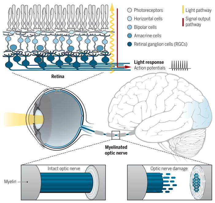
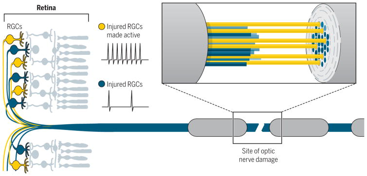
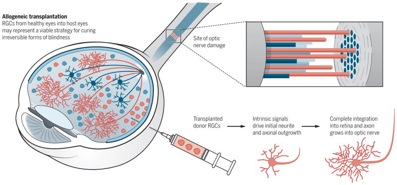
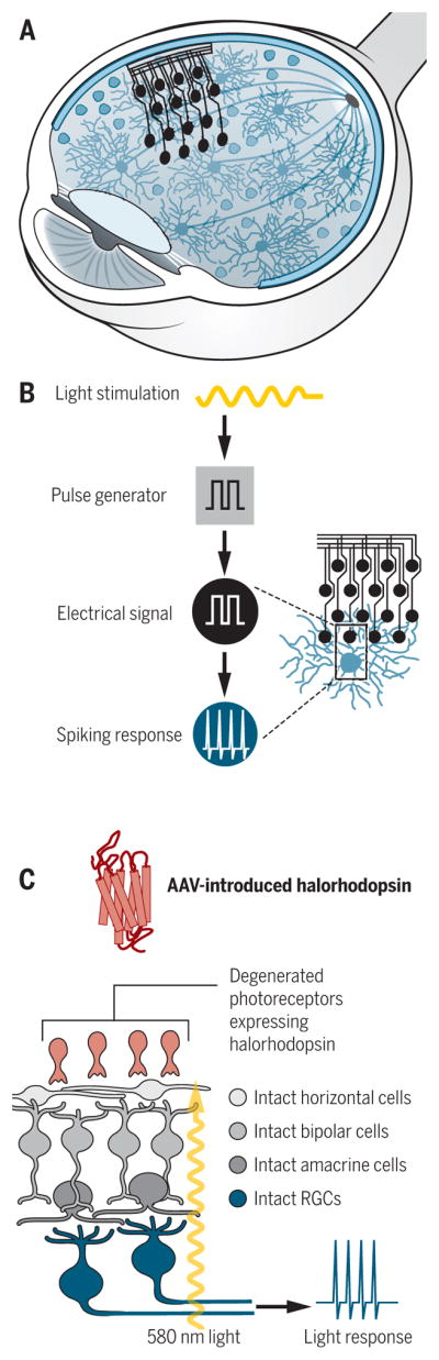
Humans are highly visual. Retinal ganglion cells (RGCs), the neurons that connect the eyes to the brain, fail to regenerate after damage, eventually leading to blindness. Here, we review research on regeneration and repair of the optic system. Intrinsic developmental growth programs can be reactivated in RGCs, neural activity can enhance RGC regeneration, and functional reformation of eye-to-brain connections is possible, even in the adult brain. Transplantation and gene therapy may serve to replace or resurrect dead or injured retinal neurons. Retinal prosthetics that can restore vision in animal models may too have practical power in the clinical setting. Functional restoration of sight in certain forms of blindness is likely to occur in human patients in the near future.


Figures

Fig. 1
Fig. 1. Visual information is transmitted from the eye to the visual centers in the brain via the optic nerve
(Top) Light reaching the retina is converted into electrical potentials that eventually cause action potentials in the ganglion cells (RGCs). (Middle) The myelinated optic nerve transmits action potentials (Bottom left) to the visual processing centers in the brain. (Bottom right) After damage, RGC axons degenerate. In the absence of therapeutic interventions, blindness ensues.
Fig. 2
Fig. 2. Electrical activity can promote RGC axon regeneration
Increasing the activity of RGCs after optic nerve damage can facilitate the repair of degenerating axons in the optic nerve (25, 33), with many extending past the site of damage into the brain and partially restoring sight in animal models (25).
Fig. 3
Fig. 3. Transplantation of RGCs to restore vision
(Left and top) Injected donor RGCs (red) differentiate and integrate into the retina after nerve damage, whereas endogenous RGCs (blue) degenerate. (Bottom right) A subset of the transplanted RGCs extend axons (red cables) down the optic nerve and ultimately reach the brain (41).
Fig. 4
Fig. 4. Retinal prostheses and virally introduced light-sensitive ion channels can potentially restore sight
(A) Electrode arrays can be surgical implanted in the retina. (B) Incoming light is converted into electrical signals by electrodes, which generate spiking in RGCs, restoring eye-to-brain communication and, potentially, sight. (C) Adeno-associated viruses (AAVs) can be used to deliver light-sensitive ion channels (halorhodopsin) to degenerated photoreceptors, allowing light stimulation at the appropriate wavelength to generate spiking in the RGCs.

Similar articles

Cited by

References

    1. Baden T, et al. Nature. 2016;529:345–350. - PMC - PubMed
    1. Quigley HA, Broman AT. Br J Ophthalmol. 2006;90:262–267. - PMC - PubMed
    1. Goldberg JL, Barres BA. Annu Rev Neurosci. 2000;23:579–612. - PubMed
    1. Case LC, Tessier-Lavigne M. Curr Biol. 2005;15:R749–R753. - PubMed
    1. Richardson PM, McGuinness UM, Aguayo AJ. Nature. 1980;284:264–265. - PubMed

Publication types

Substances