. 2017 Aug;37(8):1548-1558.
doi: 10.1161/ATVBAHA.117.309145. Epub 2017 Jun 8.

Human Blood Monocyte Subsets: A New Gating Strategy Defined Using Cell Surface Markers Identified by Mass Cytometry

Affiliations

Human Blood Monocyte Subsets: A New Gating Strategy Defined Using Cell Surface Markers Identified by Mass Cytometry

Graham D Thomas et al. Arterioscler Thromb Vasc Biol. 2017 Aug.

Abstract

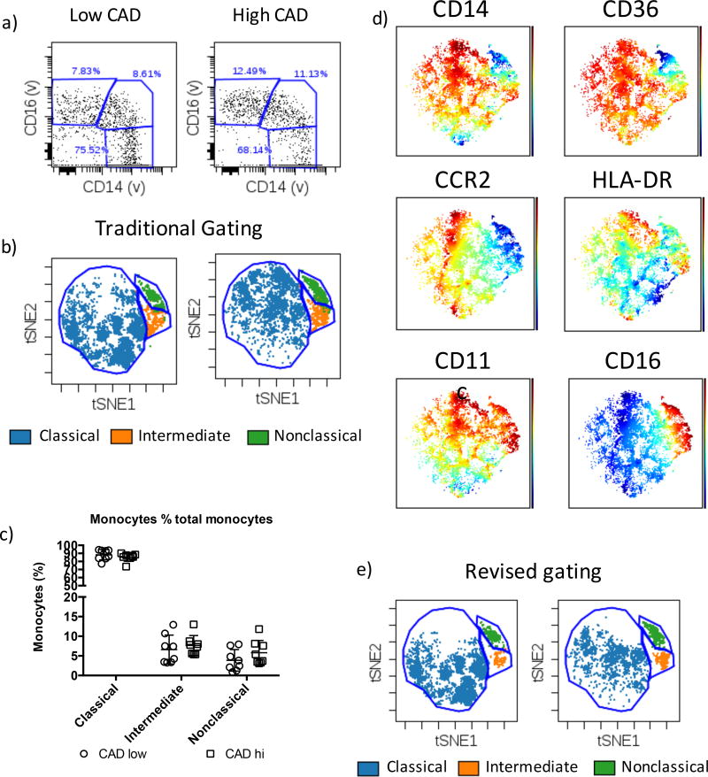
Objective: Human monocyte subsets are defined as classical (CD14++CD16-), intermediate (CD14++CD16+), and nonclassical (CD14+CD16+). Alterations in monocyte subset frequencies are associated with clinical outcomes, including cardiovascular disease, in which circulating intermediate monocytes independently predict cardiovascular events. However, delineating mechanisms of monocyte function is hampered by inconsistent results among studies.

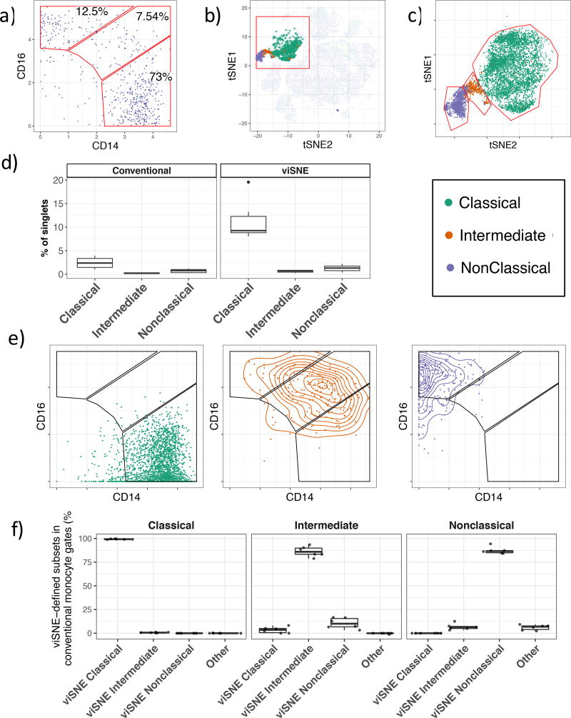
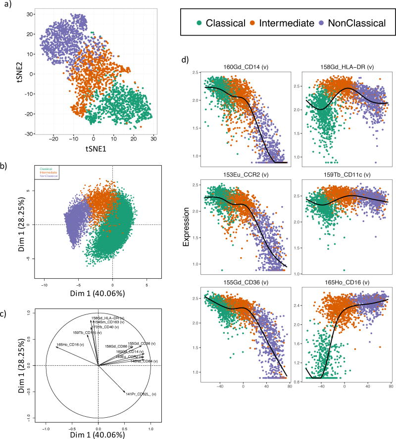
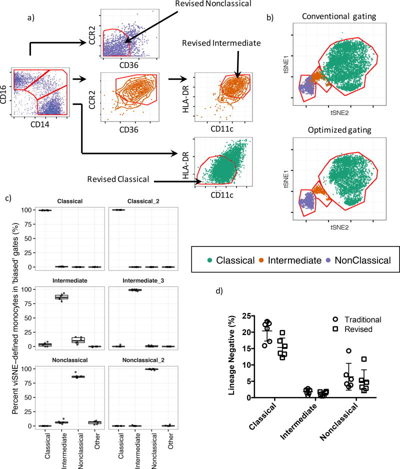
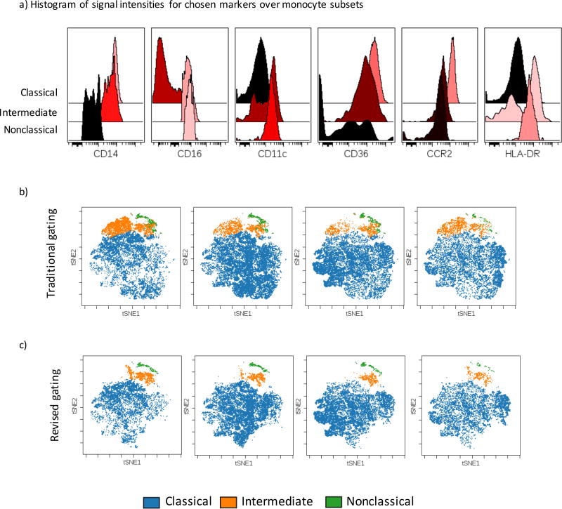
Approach and results: We use cytometry by time-of-flight mass cytometry to profile human monocytes using a panel of 36 cell surface markers. Using the dimensionality reduction approach visual interactive stochastic neighbor embedding (viSNE), we define monocytes by incorporating all cell surface markers simultaneously. Using viSNE, we find that although classical monocytes are defined with high purity using CD14 and CD16, intermediate and nonclassical monocytes defined using CD14 and CD16 alone are frequently contaminated, with average intermediate and nonclassical monocyte purity of ≈86.0% and 87.2%, respectively. To improve the monocyte purity, we devised a new gating scheme that takes advantage of the shared coexpression of cell surface markers on each subset. In addition to CD14 and CD16, CCR2, CD36, HLA-DR, and CD11c are the most informative markers that discriminate among the 3 monocyte populations. Using these additional markers as filters, our revised gating scheme increases the purity of both intermediate and nonclassical monocyte subsets to 98.8% and 99.1%, respectively. We demonstrate the use of this new gating scheme using conventional flow cytometry of peripheral blood mononuclear cells from subjects with cardiovascular disease.

Conclusions: Using cytometry by time-of-flight mass cytometry, we have identified a small panel of surface markers that can significantly improve monocyte subset identification and purity in flow cytometry. Such a revised gating scheme will be useful for clinical studies of monocyte function in human cardiovascular disease.

Keywords: atherosclerosis; flow cytometry; inflammation; monocytes.


Figures

Figure 1
Figure 1. Human monocyte gating
a) Traditional gating of classical (CD14++CD16), intermediate (CD14++, CD16+) and nonclassical (CD14+CD16+) monocytes. Monocytes are previously gated on Live, Lin (CD3, CD19, CD66b), HLADR+CD14+ cells. b) Overlay of classical, intermediate and nonclassical monocytes on a viSNE map of all PBMC acquired in our experiment. c) viSNE-based gating of monocyte subsets. Classical, intermediate and nonclassical monocytes from six individuals superimposed on a single biaxial plot showing the red region in figure 1b is shown. d) Peripheral blood monocyte frequencies defined using conventional gating or with gates drawn on the viSNE axis. e) The relative distribution of viSNE-defined classical, intermediate and non-classical monocytes within the conventional monocyte subset gates. f) Quantification of the distribution of viSNE-defined monocytes in conventional monocyte gates.
Figure 2
Figure 2. Automatic gating of human monocytes
a) viSNE analysis of human monocyte subsets. 1,000 of each classical, intermediate and nonclassical monocytes defined by viSNE were subset and clustered using all 33 conjugated antibodies. b) Principal component analysis of the three monocytes using the 11 most informative cell surface markers defined as the six top markers for each pairwise comparison between subsets based on the discrimination index. c) Principal component loading plot for figure 2b showing the relative contribution of each surface marker to each principal component. d) Isomap plots showing expression profiles of the most informative cell surface markers (defined in Table 1) during the monocyte developmental cascade.
Figure 3
Figure 3. Revised gating scheme for human monocyte subsets
a) Revised gating scheme for monocyte subsets. Conventionally gated classical monocytes are subjected to a second gating step, defined as CD11clowHLA-DRhi. Conventionally gated nonclassical monocytes are subjected to a second gating step, defined as CD36lowCCR2low. Finally conventionally gated intermediate monocytes are first subject to a gating step to filter contaminating nonclassical monocytes and are thus defined as CD36hiCCR2hi, then to a final gating step to remove contaminating classical monocytes using a CD11chiHLA-DRlow gate . b) Monocyte subsets gated using either the conventional (Figure 3a, leftmost panel) approach or our revised approach (Figure 3a, rightmost panels) overlaid on to the monocyte subset viSNE map. c) Monocyte subset purity assessed by measuring the frequency of gated cells within viSNE-defined monocyte subset gates in Figure 3b. d) Recovery of monocyte subsets gated using the additional gating steps showing only a minor loss of total monocytes with the additional gating.
Figure 4
Figure 4. Cell surface marker expression on monocyte subsets
a) Cell surface expression levels of markers in our gating scheme on Classical, Intermediate and nonclassical monocytes gated using the conventional approach showing the expected patterns of expression as determined by CyTOF. b and c) viSNE analysis of human monocytes acquired by 7 color flow cytometry, b) Traditional gating, c) revised gating strategy.
Figure 5
Figure 5. Monocyte subsets in human subjects with cardiovascular disease
a) Flow cytometry analysis showing conventional monocyte subset gating for CAD high and CAD low individuals. b) viSNE maps showing monocyte subsets gated in figure 5a and their subsequent definition using viSNE. c) Monocyte subset frequencies defined by viSNE in CAD hi and CAD low individuals. d) Rainbow plots showing the expected expression profiles of cell surface markers on the monocyte subsets. e) Revised gating strategy applied to CAD high and CAD low individuals showing improved monocyte subset classification in cardiovascular disease using our strategy.

Comment in

  • By CyTOF: Heterogeneity of Human Monocytes.
    Loke P, Niewold TB. Loke P, et al. Arterioscler Thromb Vasc Biol. 2017 Aug;37(8):1423-1424. doi: 10.1161/ATVBAHA.117.309645. Arterioscler Thromb Vasc Biol. 2017. PMID: 28747454 Free PMC article.

Similar articles

Cited by

References

    1. Ziegler-Heitbrock L, Ancuta P, Crowe S, Dalod M, Grau V, Hart DN, Leenen PJM, Liu Y-J, MacPherson G, Randolph GJ, Scherberich J, Schmitz J, Shortman K, Sozzani S, Strobl H, Zembala M, Austyn JM, Lutz MB. Nomenclature of monocytes and dendritic cells in blood. Blood. 2010;116:e74–e80. - PubMed
    1. Wong KL, Yeap WH, Tai JJY, Ong SM, Dang TM, Wong SC. The three human monocyte subsets: implications for health and disease. Immunol Res. 2012;53:41–57. - PubMed
    1. Poehlmann H, Schefold JC, Zuckermann-Becker H, Volk H-D, Meisel C. Phenotype changes and impaired function of dendritic cell subsets in patients with sepsis: a prospective observational analysis. Crit Care. 2009;13:R119. - PMC - PubMed
    1. Azeredo EL, Neves-Souza PC, Alvarenga AR, Reis SRNI, Torrentes-Carvalho A, Zagne S-MO, Nogueira RMR, Oliveira-Pinto LM, Kubelka CF. Differential regulation of toll-like receptor-2, toll-like receptor-4, CD16 and human leucocyte antigen-DR on peripheral blood monocytes during mild and severe dengue fever. Immunology. 2010;130:202–216. - PMC - PubMed
    1. Grip O, Bredberg A, Lindgren S, Henriksson G. Increased subpopulations of CD16(+) and CD56(+) blood monocytes in patients with active Crohn’s disease. Inflamm Bowel Dis. 2007;13:566–572. - PubMed

Publication types

MeSH terms