

Mitochondrial Oxidative Stress Promotes Atherosclerosis and Neutrophil Extracellular Traps in Aged Mice
- PMID: 28596373
- PMCID: PMC5535797
- DOI: 10.1161/ATVBAHA.117.309580
Mitochondrial Oxidative Stress Promotes Atherosclerosis and Neutrophil Extracellular Traps in Aged Mice
Abstract
Rationale: Mitochondrial oxidative stress (mitoOS) has been shown to be increased in various cell types in human atherosclerosis and with aging. However, the role of cell type-specific mitoOS in atherosclerosis in the setting of advanced age and the molecular mechanisms remains to be determined in vivo.
Objective: The aim of this study was to examine the role of myeloid cell mitoOS in atherosclerosis in aged mice.
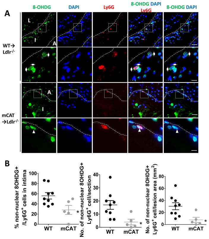
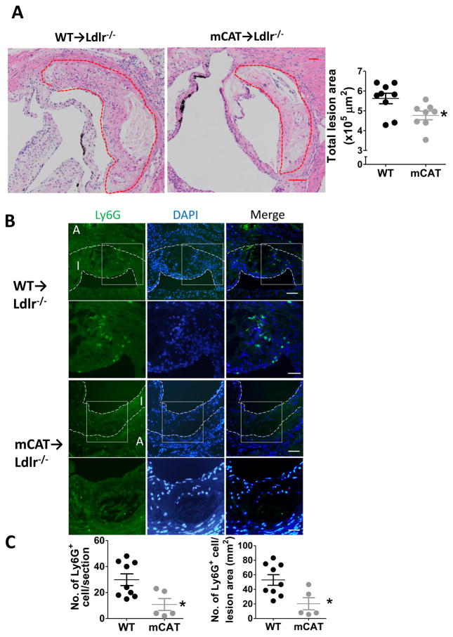
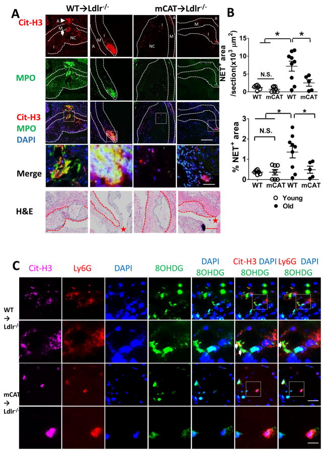
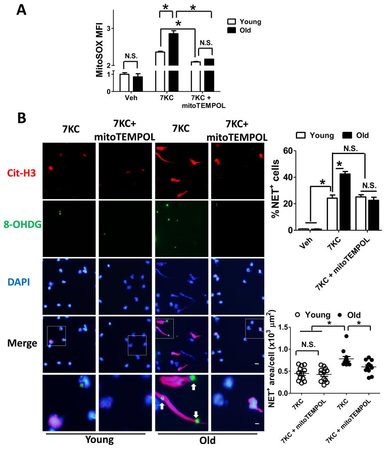
Approach and results: Lethally irradiated low-density lipoprotein receptor-deficient mice (Ldlr-/-) were reconstituted with bone marrow from either wild-type or mitochondrial catalase (mCAT) mice. mCAT transgenic mice contain ectopically expressed human catalase gene in mitochondria, which reduces mitoOS. Starting at the age of 36 weeks, mice were fed the Western-type diet for 16 weeks. We found that mitoOS in lesional myeloid cells was suppressed in aged mCAT→Ldlr-/- chimeric mice compared with aged controls, and this led to a significant reduction in aortic root atherosclerotic lesion area despite higher plasma cholesterol levels. Neutrophil extracellular traps (NETs), a proinflammatory extracellular structure that contributes to atherosclerosis progression, were significantly increased in the lesions of aged mice compared with lesions of younger mice. Aged mCAT→Ldlr-/- mice had less lesional neutrophils and decreased NETs compared with age-matched wild-type→Ldlr-/- mice, whereas young mCAT→ and wild-type→Ldlr-/- mice had comparable numbers of neutrophils and similar low levels of lesional NETs. Using cultured neutrophils, we showed that suppression of mitoOS reduced 7-ketocholesterol-induced NET release from neutrophils of aged but not younger mice.
Conclusions: MitoOS in lesional myeloid cells enhanced atherosclerosis development in aged mice, and this enhancement was associated with increased lesional NETs. Thus, mitoOS-induced NET formation is a potentially new therapeutic target to prevent atherosclerosis progression during aging.
Keywords: DNA, mitochondrial; atherosclerosis; extracellular trap; mitochondria; reactive oxygen species.
© 2017 American Heart Association, Inc.
Figures
Similar articles
-
Macrophage mitochondrial oxidative stress promotes atherosclerosis and nuclear factor-κB-mediated inflammation in macrophages.Circ Res. 2014 Jan 31;114(3):421-33. doi: 10.1161/CIRCRESAHA.114.302153. Epub 2013 Dec 2. Circ Res. 2014. PMID: 24297735 Free PMC article.
-
CalDAG-GEFI Deficiency Reduces Atherosclerotic Lesion Development in Mice.Arterioscler Thromb Vasc Biol. 2016 May;36(5):792-9. doi: 10.1161/ATVBAHA.115.306347. Epub 2016 Mar 17. Arterioscler Thromb Vasc Biol. 2016. PMID: 26988592 Free PMC article.
-
Lack of myeloid Fatp1 increases atherosclerotic lesion size in Ldlr-/- mice.Atherosclerosis. 2017 Nov;266:182-189. doi: 10.1016/j.atherosclerosis.2017.10.009. Epub 2017 Oct 7. Atherosclerosis. 2017. PMID: 29035781 Free PMC article.
-
Therapeutic Targeting of Neutrophil Extracellular Traps in Atherogenic Inflammation.Thromb Haemost. 2019 Apr;119(4):542-552. doi: 10.1055/s-0039-1678664. Epub 2019 Feb 7. Thromb Haemost. 2019. PMID: 30731493 Review.
-
Neutrophil Extracellular Traps in Atherosclerosis and Atherothrombosis.Circ Res. 2017 Feb 17;120(4):736-743. doi: 10.1161/CIRCRESAHA.116.309692. Circ Res. 2017. PMID: 28209798 Review.
Cited by
-
Efficacy of the Piperidine Nitroxide 4-MethoxyTEMPO in Ameliorating Serum Amyloid A-Mediated Vascular Inflammation.Int J Mol Sci. 2021 Apr 27;22(9):4549. doi: 10.3390/ijms22094549. Int J Mol Sci. 2021. PMID: 33925294 Free PMC article.
-
Neutrophil extracellular traps: a catalyst for atherosclerosis.Mol Cell Biochem. 2024 Dec;479(12):3213-3227. doi: 10.1007/s11010-024-04931-3. Epub 2024 Feb 24. Mol Cell Biochem. 2024. PMID: 38401035 Review.
-
High-Density Lipoprotein Suppresses Neutrophil Extracellular Traps Enhanced by Oxidized Low-Density Lipoprotein or Oxidized Phospholipids.Int J Mol Sci. 2022 Nov 13;23(22):13992. doi: 10.3390/ijms232213992. Int J Mol Sci. 2022. PMID: 36430470 Free PMC article.
-
Metformin improved oxidized low-density lipoprotein-impaired mitochondrial function and increased glucose uptake involving Akt-AS160 pathway in raw264.7 macrophages.Chin Med J (Engl). 2019 Jul 20;132(14):1713-1722. doi: 10.1097/CM9.0000000000000333. Chin Med J (Engl). 2019. PMID: 31268904 Free PMC article.
-
Atherosclerotic burden and cerebral small vessel disease: exploring the link through microvascular aging and cerebral microhemorrhages.Geroscience. 2024 Oct;46(5):5103-5132. doi: 10.1007/s11357-024-01139-7. Epub 2024 Apr 19. Geroscience. 2024. PMID: 38639833 Free PMC article. Review.
References
-
- Lee HC, Wei YH. Mitochondria and aging. Advances in experimental medicine and biology. 2012;942:311–327. - PubMed
-
- Schriner SE, Linford NJ, Martin GM, Treuting P, Ogburn CE, Emond M, Coskun PE, Ladiges W, Wolf N, Van Remmen H, Wallace DC, Rabinovitch PS. Extension of murine life span by overexpression of catalase targeted to mitochondria. Science. 2005;308:1909–1911. - PubMed
-
- Simons LA. Epidemiologic considerations in cardiovascular diseases in the elderly: International comparisons and trends. The American journal of cardiology. 1989;63:5H–8H. - PubMed
-
- Dorighello GG, Paim BA, Leite AC, Vercesi AE, Oliveira HC. Spontaneous experimental atherosclerosis in hypercholesterolemic mice advances with ageing and correlates with mitochondrial reactive oxygen species. Experimental gerontology. 2017 - PubMed
Publication types
MeSH terms
Substances
Grants and funding
LinkOut - more resources
Full Text Sources
Other Literature Sources
Medical
Molecular Biology Databases
Miscellaneous
