

ERRα Maintains Mitochondrial Oxidative Metabolism and Constitutes an Actionable Target in PGC1α-Elevated Melanomas
- PMID: 28596418
- PMCID: PMC5954239
- DOI: 10.1158/1541-7786.MCR-17-0143
ERRα Maintains Mitochondrial Oxidative Metabolism and Constitutes an Actionable Target in PGC1α-Elevated Melanomas
Abstract
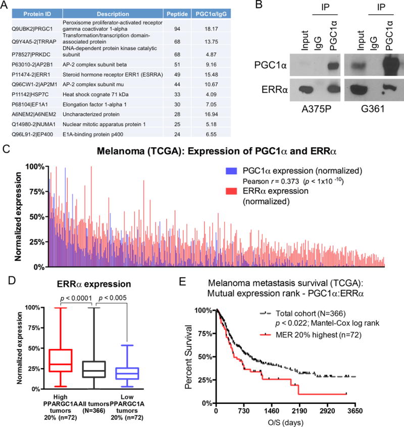
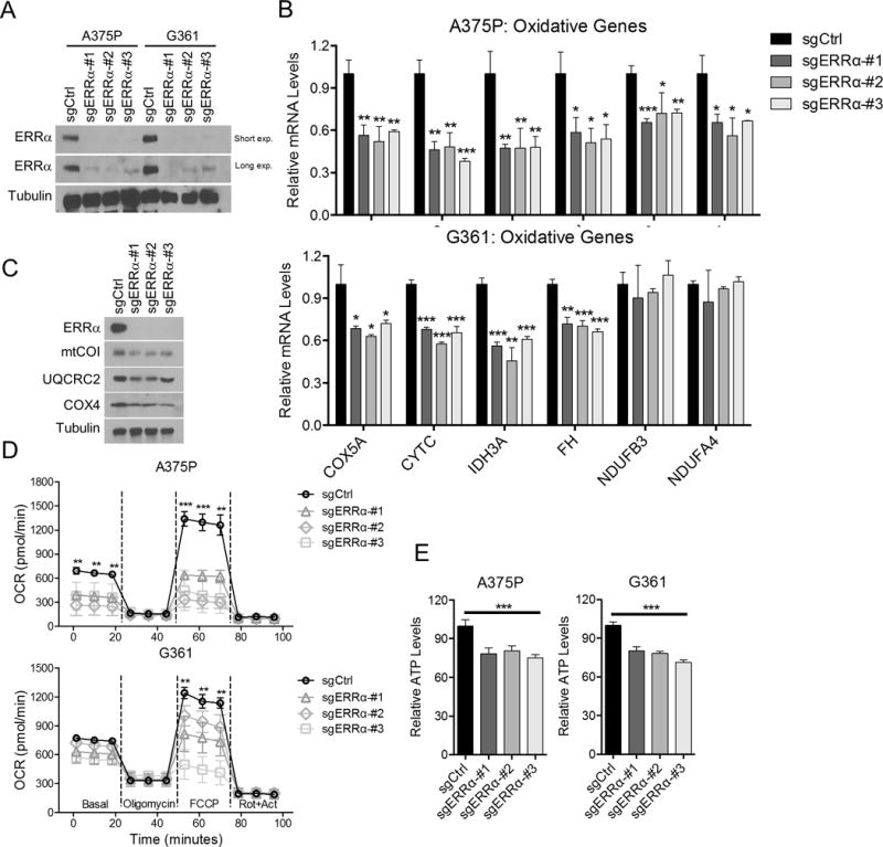
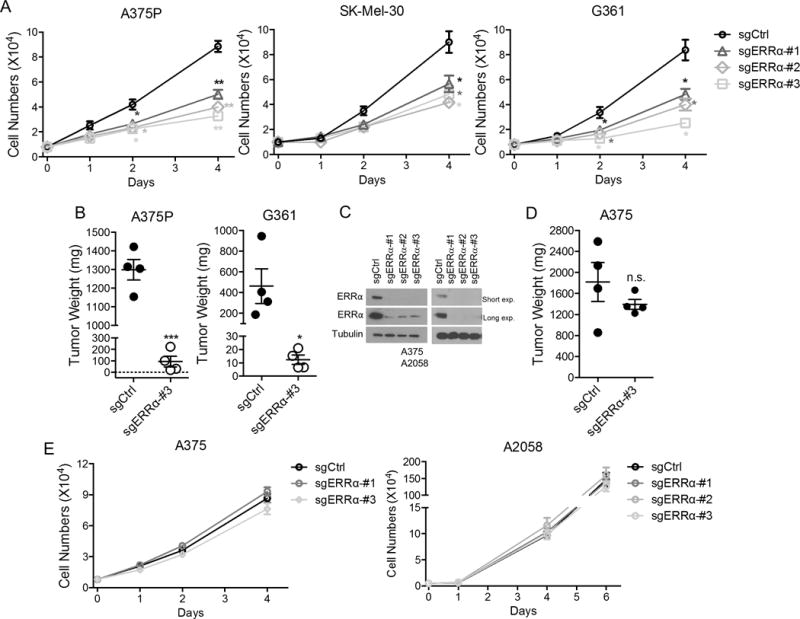
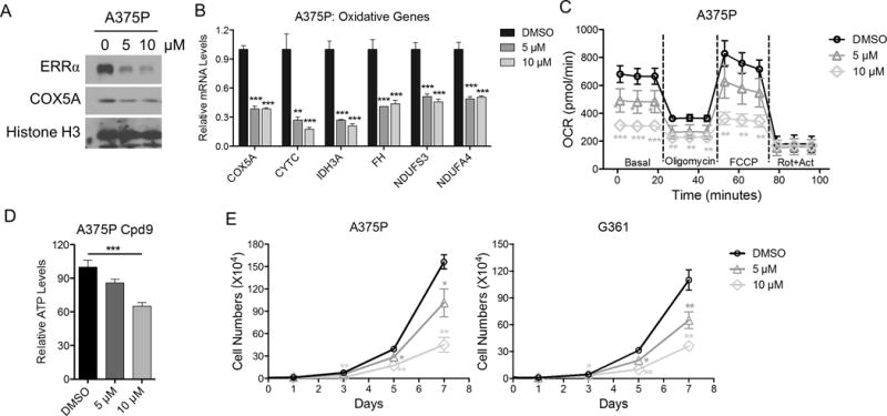
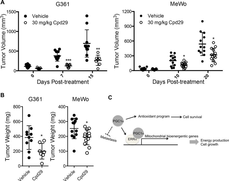
The uncontrolled growth of tumors provides metabolic dependencies that can be harnessed for therapeutic benefit. Although tumor cells exhibit these increased metabolic demands due to their rapid proliferation, these metabolic processes are general to all cells, and furthermore, targeted therapeutic intervention can provoke compensatory adaptation that alters tumors' characteristics. As an example, a subset of melanomas depends on the transcriptional coactivator PGC1α function to sustain their mitochondrial energy-dependent survival. However, selective outgrowth of resistant PGC1α-independent tumor cells becomes endowed with an augmented metastatic phenotype. To find PGC1α-dependent components that would not affect metastasis in melanomas, an unbiased proteomic analyses was performed and uncovered the orphan nuclear receptor ERRα, which supports PGC1α's control of mitochondrial energetic metabolism, but does not affect the antioxidant nor antimetastatic regulatory roles. Specifically, genetic or pharmacologic inhibition of ERRα reduces the inherent bioenergetic capacity and decreases melanoma cell growth, but without altering the invasive characteristics. Thus, within this particularly aggressive subset of melanomas, which is characterized by heighted expression of PGC1α, ERRα specifically mediates prosurvival functions and represents a tangible therapeutic target.Implications: ERRα, a druggable protein, mediates the bioenergetic effects in melanomas defined by high PGC1α expression, suggesting a rational means for therapeutic targeting of this particularly aggressive melanoma subtype. Mol Cancer Res; 15(10); 1366-75. ©2017 AACR.
©2017 American Association for Cancer Research.
Conflict of interest statement
The authors disclose no potential conflicts of interest.
Figures
Similar articles
-
A PGC1α-mediated transcriptional axis suppresses melanoma metastasis.Nature. 2016 Sep 15;537(7620):422-426. doi: 10.1038/nature19347. Epub 2016 Aug 31. Nature. 2016. PMID: 27580028 Free PMC article.
-
Targeting mitochondrial oxidative metabolism in melanoma causes metabolic compensation through glucose and glutamine utilization.Cancer Res. 2014 Jul 1;74(13):3535-45. doi: 10.1158/0008-5472.CAN-13-2893-T. Epub 2014 May 8. Cancer Res. 2014. PMID: 24812272
-
PGC1α Suppresses Prostate Cancer Cell Invasion through ERRα Transcriptional Control.Cancer Res. 2019 Dec 15;79(24):6153-6165. doi: 10.1158/0008-5472.CAN-19-1231. Epub 2019 Oct 8. Cancer Res. 2019. PMID: 31594836
-
The Role of PGC1α in Cancer Metabolism and its Therapeutic Implications.Mol Cancer Ther. 2016 May;15(5):774-82. doi: 10.1158/1535-7163.MCT-15-0621. Epub 2016 Apr 15. Mol Cancer Ther. 2016. PMID: 27197257 Review.
-
The Warburg metabolism fuels tumor metastasis.Cancer Metastasis Rev. 2019 Jun;38(1-2):157-164. doi: 10.1007/s10555-019-09794-5. Cancer Metastasis Rev. 2019. PMID: 30997670 Review.
Cited by
-
The Interaction Between Intracellular Energy Metabolism and Signaling Pathways During Osteogenesis.Front Mol Biosci. 2022 Jan 28;8:807487. doi: 10.3389/fmolb.2021.807487. eCollection 2021. Front Mol Biosci. 2022. PMID: 35155568 Free PMC article. Review.
-
The PGC-1/ERR network and its role in precision oncology.NPJ Precis Oncol. 2019 Mar 21;3:9. doi: 10.1038/s41698-019-0081-6. eCollection 2019. NPJ Precis Oncol. 2019. PMID: 30911677 Free PMC article. Review.
-
Role of PGC-1α in the proliferation and metastasis of malignant tumors.J Mol Histol. 2025 Jan 30;56(2):77. doi: 10.1007/s10735-025-10360-3. J Mol Histol. 2025. PMID: 39881043 Review.
-
In-depth proteomic profiling captures subtype-specific features of craniopharyngiomas.Sci Rep. 2021 Oct 27;11(1):21206. doi: 10.1038/s41598-021-00483-4. Sci Rep. 2021. PMID: 34707096 Free PMC article.
-
ERRα is an aggressive factor in lung adenocarcinoma indicating poor prognostic outcomes.Cancer Manag Res. 2019 Sep 2;11:8111-8123. doi: 10.2147/CMAR.S204732. eCollection 2019. Cancer Manag Res. 2019. PMID: 31564971 Free PMC article.
References
Publication types
MeSH terms
Substances
Grants and funding
LinkOut - more resources
Full Text Sources
Other Literature Sources
Medical
Research Materials
