. 2017 Jul 18;8(29):46769-46780.
doi: 10.18632/oncotarget.18151.

Rumen-derived lipopolysaccharide provoked inflammatory injury in the liver of dairy cows fed a high-concentrate diet

Affiliations

Rumen-derived lipopolysaccharide provoked inflammatory injury in the liver of dairy cows fed a high-concentrate diet

Junfei Guo et al. Oncotarget. .

Abstract

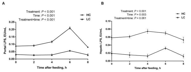
Rumen-derived lipopolysaccharide (LPS) is translocated from the rumen into the bloodstream when subacute ruminal acidosis (SARA) occurs following long-term feeding with a high-concentrate (HC) diet in dairy cows. The objective of this study was to investigate the mechanism of inflammatory responses in the liver caused by HC diet feeding. We found that SARA was induced in dairy cows when rumen pH below 5.6 lasted for at least 3 h/d with HC diet feeding. Also, the LPS levels in the portal and hepatic veins were increased significantly and hepatocytes were impaired as well as the liver function was inhibited during SARA condition. Meanwhile, the mRNA expression of immune genes including TNF receptor associated factor 6 (TRAF6), nuclear factor-kappa B (NF-κB), p38 mitogen-activated protein kinase (MAPK), extracellular regulated protein kinases (ERK) MAPK, Interleukin-1 (IL-1) and serum amyloid A (SAA) in the liver were significantly increased in SARA cows. Moreover, the phosphorylation level of NF-κB p65 and p38 MAPK proteins in the liver and the concentration of Tumor Necrosis Factor (TNF-α), Interleukin-1β (IL-1β) and Interleukin-6 (IL-6) in peripheral blood were obviously increased under SARA condition. In conclusion, the inflammatory injury in the liver caused by LPS that traveled from the digestive tract to the liver through the portal vein after feeding with a HC diet.

Keywords: Immune response; Immunity; Immunology and Microbiology Section; dairy cows; inflammatory injury; lipopolysaccharide; liver; subacute ruminal acidosis.


Conflict of interest statement

CONFLICTS OF INTEREST

The authors declare no conflicts of interest.

Figures

Figure 1
Figure 1. pH value of rumen fluid of high-concentration (HC) and low-concentrate (LC) diet
Rumen pH value was measured at different time points post feeding. Rumen pH decreased dramatically in HC group compared to LC group (mean ± SEM, n = 6). Differences between two groups were considered as significant when P < 0.05.
Figure 2
Figure 2. The concentration of LPS in portal and hepatic veins at different time points post feeding in the liver of dairy cows from high-concentrate (HC) and low-concentrate (LC) diet
A. LPS concentration in portal vein (mean ± SEM, n = 6). B. LPS concentration in hepatic vein (mean ± SEM, n = 6). LPS concentration in HC group was much higher than LC group. Difference was considered as significant between two groups when P < 0.05.
Figure 3
Figure 3. The effect of high-concentrate (HC) diet on hepatocytes histopathologic changes in dairy cows
Representative photomicrographs with hematoxylin and eosin staining. HC: High-concentrate diet. LC: Low-concentrate diet. A., B. and C. LC group. D., E. and F. HC group. G. Hepatocytes injury score. All the sections were observed at 100, 200 or 400 × magnification. GN: Glycogenated nuclei. ICI: Inflammatory cells infiltrate. ICIB: Liver cells injury ballooning. The thin arrow indicates GN damages; thick right arrow in F indicates ICIB damages while in C indicates no disruption; thick left arrow indicates inflammatory cells infiltrate. The damage score are expressed as the mean ± SEM and asterisks indicate the significant difference between two groups (**P < 0.01).
Figure 4
Figure 4. The mRNA relative expression of immune genes in the liver of dairy cows from high-concentrate (HC) and low-concentrate (LC) diet detected by Real-Time qPCR
A. Immune genes involved in inflammatory signaling pathway. The expression of TRAF6, NF-κB-p38 MAPK and ERK MAPK in the liver were increased in HC group compare to LC group. B. Pro- and anti-inflammatory cytokines and acute phase proteins. The expression of IL-1βand SAA in the liver were obviously higher in HC group than LC group. The data are expressed as the mean ± SEM and asterisks indicate the differences between two groups (*P < 0.05, n = 6).
Figure 5
Figure 5. NF-κB p65
A., p38 B. MAPK and ERK MAPK C. phosphorylation levels in the liver of dairy cows from high-concentrate (HC) and low-concentrate (LC) diet. The phosphorylation level of NF-κB and p38 MAPK in the liver were significantly higher in HC group than LC group. The data are expressed as the mean ± SEM, and the asterisks indicate the significant differences between two groups (*P < 0.05, n = 6).
Figure 6
Figure 6. The concentrations of IL-1β
A., IL-6 B., and TNF-α C. in peripheral blood of dairy cows from high-concentrate (HC) and low-concentrate (LC) diet. All of three pro-inflammatory cytokines were increased obviously in peripheral blood in HC group compared with LC group. The data are expressed as the mean ± SEM, and asterisks indicate the significant differences between two groups (*P < 0.05, n = 6).
Figure 7
Figure 7. The route of LPS transported to the liver in dairy cows
LPS in gastrointestinal tract results from high-concentrate diet was translocated into the liver via portal vein. Then LPS combined with LBP and transported to the hepatocytes surface with the help of CD14, and recognized by TLR4. Subsequently, the complex combined with TRIF or myeloid differentiation factor 88 (MyD88) and activates TRAF6, followed by the activation of Transforming growth factor beta-activated kinase 1 (TAK1). On the one hand, TAK1 activate inhibitor of nuclear factor kappa-B kinases (IKKs) so that NF-κB could be able to escape from IKKs and translocated into the nucleus to stimulate the pro-inflammatory cytokines such as IL-1 and IL-6. On the other hand, TAK1 activate the MAPK to stimulate the pro-inflammatory cytokines production. Those pro-inflammatory cytokines released into the blood circulation and trigger the synthesis of APPs in the liver. As a result, acute phase responses in the body are induced.

Similar articles

Cited by

References

    1. Krause KM, Oetzel GR. Understanding and preventing subacute ruminal acidosis in dairy herds: A review. Anim Feed Sci Tech. 2006;126:215–36. doi: 10.1016/j.anifeedsci.2005.08.004. - DOI
    1. Gozho G, Plaizier J, Krause D, Kennedy A, Wittenberg K. Subacute ruminal acidosis induces ruminal lipopolysaccharide endotoxin release and triggers an inflammatory response. J Dairy Sci. 2005;88:1399–403. doi: 10.3168/jds.S0022-0302(05)72807-1. - DOI - PubMed
    1. Gozho G, Krause D, Plaizier J. Rumen lipopolysaccharide and inflammation during grain adaptation and subacute ruminal acidosis in steers. J Dairy Sci. 2006;89:4404–13. doi: 10.3168/jds.S0022-0302(06)72487-0. - DOI - PubMed
    1. Plaizier J, Krause D, Gozho G, McBride B. Subacute ruminal acidosis in dairy cows: The physiological causes, incidence and consequences. Vet J. 2008;176:21–31. doi: 10.1016/j.tvjl.2007.12.016. - DOI - PubMed
    1. Nocek JE. Bovine acidosis: Implications on laminitis. J Dairy Sci. 1997;80:1005–28. doi: 10.3168/jds.S0022-0302(97)76026-0. - DOI - PubMed

MeSH terms

LinkOut - more resources